A Long Journey to the Truth: Primary Cardiac Lymphoma with Various Arrhythmias from Ventricular Tachycardia to Atrial Flutter
- Affiliations
-
- 1Division of Cardiology, Department of Internal Medicine, Seoul St. Mary's Hospital, College of Medicine, The Catholic University of Korea, Seoul, Korea. oys@catholic.ac.kr
- 2Division of Cardiology, Department of Internal Medicine, Dongguk University Ilsan Hospital, Goyang, Korea.
- KMID: 2471290
- DOI: http://doi.org/10.4070/kcj.2019.0298
Abstract
- No abstract available.
Figure
-
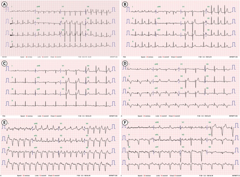
Figure 1 Twelve-lead ECGs. (A) ECG at the time of the first hospital visit showed 1st degree AV block (PR interval 360 ms). (B) Mobitz type I second-degree AV block was also recorded. (C) In the 3rd year after the first visit, T inversion began to be observed. (D) In the 4th year after the first visit, deep T-inversion proceeded in inferior leads and lateral precordial leads. (E) In the 5th year after the first visit, intractable ventricular tachycardia developed. (F) In the 7th year, after steroid pulse therapy for sarcoidosis, a typical atrial flutter occurred.AV = atrioventricular; ECG = electrocardiogram.

Figure 2 Images of transthoracic echocardiography. (A) Transthoracic echocardiogram at the time of the first hospital visit showed normal findings. (B) In the 4th year after the first visit, it indicated hypertrophied posterior wall thickness with hyperechoic myocardium. (C) In the 5th year after the first visit, unusual pattern of LV inferolateral wall hypertrophy was shown. (D) In the 7th year after the first visit, more progression of asymmetric LV hypertrophy and a huge mass in RA cavity with near-total obstruction of flow across tricuspid valve were observed. (E) Nine months after chemotherapy, the huge mass involving interatrial septum disappeared and LV wall thickness was nearly normalized.Ao = aorta; LA = left atrial; LV = left ventricular; RA = right atrial; RV = right ventricular.
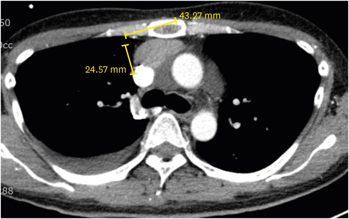
Figure 3 Chest computed tomography in the 7th year after the first hospital visit revealed anterior mediastinal lymph node enlargement.
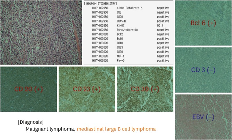
Figure 4 Anterior mediastinal lymph node excision biopsy showed a malignant large B cell lymphoma.Bcl = B-cell lymphoma; CD = cluster of differentiation; EBV = Epstein-Barr virus.

Figure 5 Images of positron emission tomography-computed tomography. (A) In the 7th year after the first visit, intense focal FDG uptakes were observed in the LA and LV wall. (B) Six months after starting rituximab, cyclophosphamide, doxorubicin, vincristine, and prednisolone chemotherapy, the FDG uptake in the LV wall decreased significantly.FDG = fluoro-deolyglucose; LA = left atrial; LV = left ventricular.
Cited by 1 articles
-
The Long Journey of Cardiac Lymphoma Follow-up
Joseph C. Lee, Yi-Tung Tom Huang, Yu-Ting Huang, Jia Wen Chong, William W. Chik
Korean Circ J. 2020;50(6):533-534. doi: 10.4070/kcj.2020.0101.
Reference
-
1. Jeudy J, Kirsch J, Tavora F, et al. From the radiologic pathology archives: cardiac lymphoma: radiologic-pathologic correlation. Radiographics. 2012; 32:1369–1380. PMID: 22977025.2. Mendelson L, Hsu E, Chung H, Hsu A. Primary cardiac lymphoma: importance of tissue diagnosis. Case Rep Hematol. 2018; 6192452. PMID: 30147970.3. Petrich A, Cho SI, Billett H. Primary cardiac lymphoma: an analysis of presentation, treatment, and outcome patterns. Cancer. 2011; 117:581–589. PMID: 20922788.4. Tai CJ, Wang WS, Chung MT, et al. Complete atrio-ventricular block as a major clinical presentation of the primary cardiac lymphoma: a case report. Jpn J Clin Oncol. 2001; 31:217–220. PMID: 11450997.
- Full Text Links
-
- Actions
-
Cited
- CITED
-
- Close
- Share
-
- Similar articles
-
- Differential Diagnosis of Supraventricular Tachycardia
- Perimortem cesarean section in a pregnant woman with flecainide-induced ventricular tachycardia: A case report
- A Study on Propranolol as Anti-Arrhythmic Agent
- Management of Atrial Flutter
- Atrial flutter associated with high pressure pneumoperitoneum during laparoscopic gastrectomy: A case report
