A Novel Pancreatic Imaging Window for Stabilized Longitudinal In Vivo Observation of Pancreatic Islets in Murine Model
- Affiliations
-
- 1Department of Emergency Medicine, Seoul National University Bundang Hospital, Seoul National University College of Medicine, Seongnam, Korea.
- 2Graduate School of Medical Science and Engineering, Korea Advanced Institute of Science and Technology (KAIST), Daejeon, Korea. pilhan.kim@kaist.ac.kr
- 3Graduate School of Nanoscience and Technology, Korea Advanced Institute of Science and Technology (KAIST), Daejeon, Korea.
- 4KI for Health Science and Technology (KIHST), Korea Advanced Institute of Science and Technology (KAIST), Daejeon, Korea.
- KMID: 2470967
- DOI: http://doi.org/10.4093/dmj.2018.0268
Abstract
- Longitudinal imaging of murine pancreas is technically challenging due to the mechanical softness of the tissue influenced by peristalsis. Here, we report a novel pancreatic imaging window for long-term stabilized cellular-level observation of the islets in the pancreas in vivo. By spatially separating the pancreas from the bowel movement and physiologic respiration with a metal plate integrated in the imaging window, we successfully tracked the pancreatic islets up to three weeks and visualized the dumbbell-shape transformation from the single islet. This window can be a useful tool for long-term cellular-level visualization of the microstructure in the pancreas.
Keyword
Figure
-

Fig. 1 A novel murine pancreatic window for long-term stabilized imaging of islets in vivo. (A) Schematic diagram of the pancreas arrangement in the pancreatic imaging window. (B) Photograph of the pancreatic imaging window. (C) Photograph of the application of the pancreatic imaging window in the mouse on an imaging stage with a window holder. (D) Wide-area mosaic and magnified image of the islet in the pancreas and of the vasculature in the mouse insulin 1 promoter (MIP)-green fluorescent protein (GFP) mouse. Scale bar, 1 mm (mosaic), 100 µm (magnified). (E) Comparison of the long-term tissue stability in islet imaging using the abdominal imaging window and pancreas imaging window. Each arrowhead with different colors indicates the same islets. Scale bar, 500 µm.
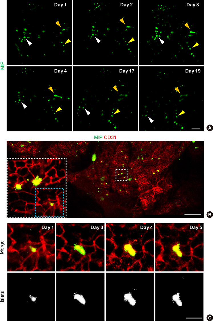
Fig. 2 Longitudinal imaging of the islets and the dumbbell-shape transformation of the islets. (A) Longitudinal imaging of the islets for 19 days in mouse insulin 1 promoter (MIP)-green fluorescent protein (GFP) mouse. Each arrowhead with different colors indicates the same islets. Scale bar, 100 µm. (B) Dumbbell-shape transformation of the islets in the pancreas. Wide-area mosaic imaging and the magnified view. Scale bar, 500 µm. (C) Magnified field of view over a period of 5 days, showing the dumbbell-shape transformation initiated from a single islet in the pancreas. Scale bar, 50 µm.
Reference
-
1. Ritsma L, Steller EJ, Beerling E, Loomans CJ, Zomer A, Gerlach C, Vrisekoop N, Seinstra D, van Gurp L, Schafer R, Raats DA, de Graaff A, Schumacher TN, de Koning EJ, Rinkes IH, Kranenburg O, van Rheenen J. Intravital microscopy through an abdominal imaging window reveals a pre-micrometastasis stage during liver metastasis. Sci Transl Med. 2012; 4:158ra145.
Article2. Ritsma L, Steller EJ, Ellenbroek SI, Kranenburg O, Borel Rinkes IH, van Rheenen J. Surgical implantation of an abdominal imaging window for intravital microscopy. Nat Protoc. 2013; 8:583–594.
Article3. Ritsma L, Ellenbroek SIJ, Zomer A, Snippert HJ, de Sauvage FJ, Simons BD, Clevers H, van Rheenen J. Intestinal crypt homeostasis revealed at single-stem-cell level by in vivo live imaging. Nature. 2014; 507:362–365.
Article4. Dolensek J, Rupnik MS, Stozer A. Structural similarities and differences between the human and the mouse pancreas. Islets. 2015; 7:e1024405.
Article5. Pittet MJ, Weissleder R. Intravital imaging. Cell. 2011; 147:983–991.
Article6. Woodward SC, Herrmann JB, Cameron JL, Brandes G, Pulaski EJ, Leonard F. Histotoxicity of cyanoacrylate tissue adhesive in the rat. Ann Surg. 1965; 162:113–122.
Article7. Kim P, Puoris'haag M, Cote D, Lin CP, Yun SH. In vivo confocal and multiphoton microendoscopy. J Biomed Opt. 2008; 13:010501.
Article8. Kim P, Chung E, Yamashita H, Hung KE, Mizoguchi A, Kucherlapati R, Fukumura D, Jain RK, Yun SH. In vivo wide-area cellular imaging by side-view endomicroscopy. Nat Methods. 2010; 7:303–305.
Article9. Park I, Choe K, Seo H, Hwang Y, Song E, Ahn J, Hwan Jo Y, Kim P. Intravital imaging of a pulmonary endothelial surface layer in a murine sepsis model. Biomed Opt Express. 2018; 9:2383–2393.
Article10. Hara M, Wang X, Kawamura T, Bindokas VP, Dizon RF, Alcoser SY, Magnuson MA, Bell GI. Transgenic mice with green fluorescent protein-labeled pancreatic beta-cells. Am J Physiol Endocrinol Metab. 2003; 284:E177–E183.11. Jo J, Kilimnik G, Kim A, Guo C, Periwal V, Hara M. Formation of pancreatic islets involves coordinated expansion of small islets and fission of large interconnected islet-like structures. Biophys J. 2011; 101:565–574.
Article12. Jo J, Hara M, Ahlgren U, Sorenson R, Periwal V. Mathematical models of pancreatic islet size distributions. Islets. 2012; 4:10–19.
Article13. van Gurp L, Loomans CJM, van Krieken PP, Dharmadhikari G, Jansen E, Ringnalda FCAS, Beerling E, van Rheenen J, de Koning EJP. Sequential intravital imaging reveals in vivo dynamics of pancreatic tissue transplanted under the kidney capsule in mice. Diabetologia. 2016; 59:2387–2392.
Article14. Lee EM, Park I, Lee YJ, You YH, Kim JW, Kim MJ, Ahn YB, Kim P, Ko SH. Effect of resveratrol treatment on graft revascularization after islet transplantation in streptozotocin-induced diabetic mice. Islets. 2018; 10:25–39.
Article15. Seymour PA, Bennett WR, Slack JM. Fission of pancreatic islets during postnatal growth of the mouse. J Anat. 2004; 204:103–116.
Article16. Jain RK. Antiangiogenesis strategies revisited: from starving tumors to alleviating hypoxia. Cancer Cell. 2014; 26:605–622.
Article17. Herreros-Villanueva M, Hijona E, Cosme A, Bujanda L. Mouse models of pancreatic cancer. World J Gastroenterol. 2012; 18:1286–1294.
Article
- Full Text Links
-
- Actions
-
Cited
- CITED
-
- Close
- Share
-
- Similar articles
-
- Comparative study of endocrine cells in the principal pancreatic islets of two teleosts, Silurus asotus (Siluridae) and Siniperca scherzeri (Centropomidae)
- Immunohistochemical Study of the Pancreatic Endocrine Cells in the BALB/c mice: An Unique Distributional Pattern of Glucagon
- Immunocytochemical Expression of Amylin in Pancreatic Islets of Man, Rabbit and Guinea Pig
- Protective Effect of Human Mesenchymal Stem Cells on the Survival of Pancreatic Islets
- An immunohistochemical study on the pancreatic islets cells of the Mongolian gerbils, Meriones unguiculatus
