A Congenital Annular Pericardial Fibrous Band Diagnosed at Fetal Life
- Affiliations
-
- 1Department of Pediatrics, Seoul St. Mary's Hospital, College of Medicine, The Catholic University of Korea, Seoul, Korea. jaeyounglee@catholic.ac.kr
- KMID: 2470915
- DOI: http://doi.org/10.4070/kcj.2019.0274
Abstract
- No abstract available.
Figure
-
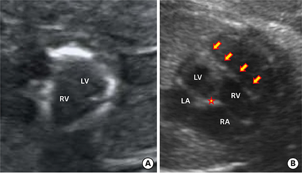
Figure 1 Fetal echocardiography findings. (A) A short-axis view showing an unusual white ring-like structure encircling both mid-ventricular cavities. (B) A 4-chamber view showing a band-like structure crossing diaphragmatic surface of both ventricles (arrows). The star (★) indicates the crux cordis. LA = left atrium; LV = left ventricle; RA = right atrium; RV = right ventricle.
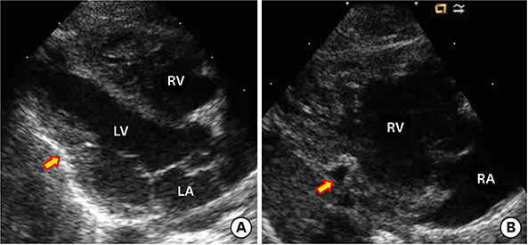
Figure 2 Parasternal long-axis views showing invaginations of pericardium (arrows) into diaphragmatic surfaces of LV (A) and RV (B). LA = left atrium; LV = left ventricle; RA = right atrium; RV = right ventricle.
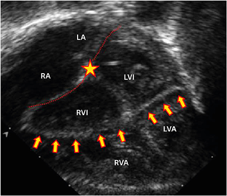
Figure 3 A modified apical 4-chamber view showing a band-like structure crossing diaphragmatic surface of both ventricles (arrows). The star (★) indicates the crux cordis and dotted lines indicate atrioventricular junctions. LA = left atrium; LVA = apical part of left ventricle; LVI = inlet part of left ventricle; RA = right atrium; RVA = apical part of right ventricle; RVI = inlet part of right ventricle.
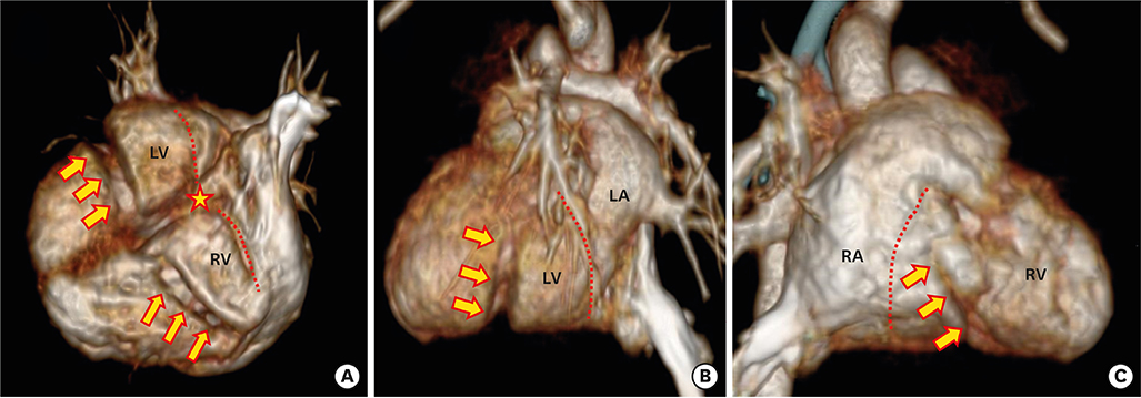
Figure 4 Three-dimensional cardiac computed tomography showing indentations (arrows) of diaphragmatic surface of both ventricles (A), lateral wall of LV (B) and lateral wall of RV (C). The star (★) indicates the crux cordis and dotted lines indicate atrioventricular groove or junctions. LA = left atrium; LV = left ventricle; RA = right atrium; RV = right ventricle.
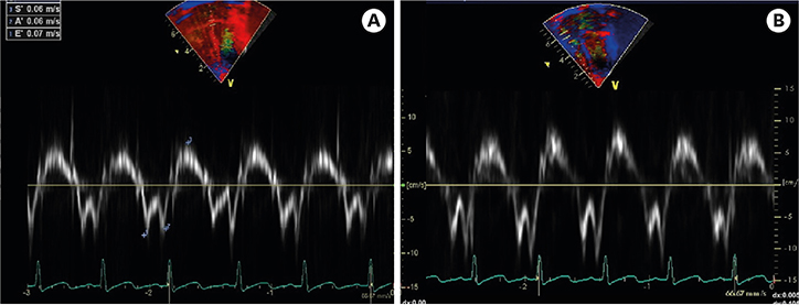
Figure 5 Tissue Doppler images showing decreased early diastolic (E′) velocities of mitral (A) and tricuspid annuli (B), suggesting an early stage of ventricular diastolic dysfunction.
Reference
-
1. Parmar YJ, Shah AB, Poon M, Kronzon I. Congenital abnormalities of the pericardium. Cardiol Clin. 2017; 35:601–614.
Article2. Gautam MP, Gautam S, Sogunuru G, Subramanyam G. Constrictive pericarditis with a calcified pericardial band at the level of left ventricle causing mid-ventricular obstruction. BMJ Case Rep. 2012; 2012:bcr0920114743.
Article3. Karakus A, Ari H, Camci S, Ari S, Tutuncu A, Melek M. Hourglass-shaped right ventricle and localized constrictive pericarditis. Echocardiography. 2017; 34:320–321.
Article4. Adler Y, Charron P. The 2015 ESC guidelines on the diagnosis and management of pericardial diseases. Eur Heart J. 2015; 36:2873–2874.
- Full Text Links
-
- Actions
-
Cited
- CITED
-
- Close
- Share
-
- Similar articles
-
- Annular Pancreas with Meckel’s Diverticulum and Ladd’s Band in Neonate
- Two Cases of Firotic Band
- Extension Contracture of Both Hip Joints Secondary to Congenital Fibrous Bands of Both Gluteus Maximus Muscles: Case Report
- Unilateral Hydronephrosis Caused by Fibrous Band Around The Ureteropelvic Junction
- A Case of Amniotic Band Syndrome Associated with Gastroschisis
