J Breast Cancer.
2019 Dec;22(4):635-640. 10.4048/jbc.2019.22.e58.
Intraoperative Specimen Mammography for Margin Assessment in Breast-Conserving Surgery
- Affiliations
-
- 1Department of Surgery, Ajou University Hospital, Suwon, Korea. smartblade@gmail.com
- 2Department of Radiology, Ajou University Hospital, Suwon, Korea.
- KMID: 2470899
- DOI: http://doi.org/10.4048/jbc.2019.22.e58
Abstract
- The purpose of this study aimed to determine whether intraoperative specimen mammography is an effective margin assessment method in Asian women. Thus, 182 patients who underwent breast-conserving surgery (BCS) were evaluated. After wide excision, intraoperative specimen mammography was used to assess margin adequacy. The control group comprised 84 patients who underwent BCS and were evaluated for margin of frozen section during surgery. 61.6% patients had dense breasts and 85.7% of dense breasts could margin assess by intraoperative specimen mammography. There were no significant differences in the incidence of extremely close margins (p = 0.421) and second operation (p = 0.252) between both groups. Significant correlations were found between radiological and histological margins (R² = 0.222, p < 0.05). The frozen section analysis group had longer operative time than the specimen mammography group. The study results show that intraoperative specimen mammography of breast lesions in BCS is useful in identifying margin clearance.
Keyword
MeSH Terms
Figure
-
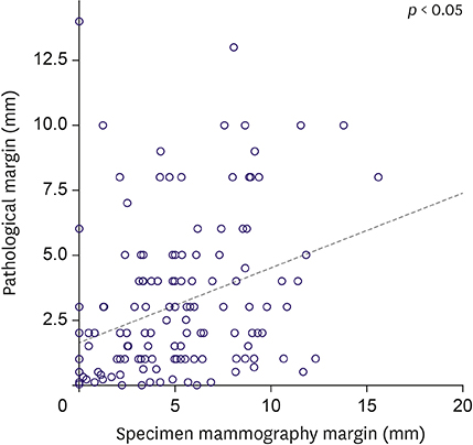
Figure 1 Correlation between the intraoperative specimen radiological margins and the histological margins nearest distance lesion (R2 = 0.222, p < 0.05).
Reference
-
1. Weber WP, Engelberger S, Viehl CT, Zanetti-Dallenbach R, Kuster S, Dirnhofer S, et al. Accuracy of frozen section analysis versus specimen radiography during breast-conserving surgery for nonpalpable lesions. World J Surg. 2008; 32:2599–2606.
Article2. Bimston DN, Bebb GG, Wagman LD. Is specimen mammography beneficial? Arch Surg. 2000; 135:1083–1086.
Article3. Kim SH, Kim MH, Oh KK. Analysis and comparison of breast density according to age on mammogram between Korea and western women. J Korean Radiol Soc. 2000; 42:1009–1014.
Article4. American College of Radiology. ACR BI-RADS® Atlas. 5th edition. Reston (VA): American College of Radiology;2013.5. Gajdos C, Tartter PI, Bleiweiss IJ, Hermann G, de Csepel J, Estabrook A, et al. Mammographic appearance of nonpalpable breast cancer reflects pathologic characteristics. Ann Surg. 2002; 235:246–251.
Article6. Papa MZ, Klein E, Davidson B, Karni T, Sperber F, Koller M, et al. The effect of anesthesia type on needle localization breast biopsy: another point of view. Am J Surg. 1996; 171:242–243.
Article7. Mullen R, Macaskill EJ, Khalil A, Elseedawy E, Brown DC, Lee AC, et al. Involved anterior margins after breast conserving surgery: is re-excision required? Eur J Surg Oncol. 2012; 38:302–306.
Article8. Jung W, Kang E, Kim SM, Kim D, Hwang Y, Sun Y, et al. Factors associated with re-excision after breast-conserving surgery for early-stage breast cancer. J Breast Cancer. 2012; 15:412–419.
Article9. Esbona K, Li Z, Wilke LG. Intraoperative imprint cytology and frozen section pathology for margin assessment in breast conservation surgery: a systematic review. Ann Surg Oncol. 2012; 19:3236–3245.
Article10. Osborn JB, Keeney GL, Jakub JW, Degnim AC, Boughey JC. Cost-effectiveness analysis of routine frozen-section analysis of breast margins compared with reoperation for positive margins. Ann Surg Oncol. 2011; 18:3204–3209.
Article11. Riedl O, Fitzal F, Mader N, Dubsky P, Rudas M, Mittlboeck M, et al. Intraoperative frozen section analysis for breast-conserving therapy in 1016 patients with breast cancer. Eur J Surg Oncol. 2009; 35:264–270.
Article12. Cendán JC, Coco D, Copeland EM 3rd. Accuracy of intraoperative frozen-section analysis of breast cancer lumpectomy-bed margins. J Am Coll Surg. 2005; 201:194–198.
Article13. Ahmed M, van Hemelrijck M, Douek M. Systematic review of radioguided versus wire-guided localization in the treatment of non-palpable breast cancers. Breast Cancer Res Treat. 2013; 140:241–252.
Article14. Bathla L, Harris A, Davey M, Sharma P, Silva E. High resolution intra-operative two-dimensional specimen mammography and its impact on second operation for re-excision of positive margins at final pathology after breast conservation surgery. Am J Surg. 2011; 202:387–394.
Article15. Layfield DM, May DJ, Cutress RI, Richardson C, Agrawal A, Wise M, et al. The effect of introducing an in-theatre intra-operative specimen radiography (IOSR) system on the management of palpable breast cancer within a single unit. Breast. 2012; 21:459–463.
Article16. Allweis TM, Kaufman Z, Lelcuk S, Pappo I, Karni T, Schneebaum S, et al. A prospective, randomized, controlled, multicenter study of a real-time, intraoperative probe for positive margin detection in breast-conserving surgery. Am J Surg. 2008; 196:483–489.
Article17. Mazouni C, Rouzier R, Balleyguier C, Sideris L, Rochard F, Delaloge S, et al. Specimen radiography as predictor of resection margin status in non-palpable breast lesions. Clin Radiol. 2006; 61:789–796.
Article18. Martin LJ, Boyd NF. Mammographic density. Potential mechanisms of breast cancer risk associated with mammographic density: hypotheses based on epidemiological evidence. Breast Cancer Res. 2008; 10:201.
Article19. Habel LA, Capra AM, Oestreicher N, Greendale GA, Cauley JA, Bromberger J, et al. Mammographic density in a multiethnic cohort. Menopause. 2007; 14:891–899.
Article20. El-Bastawissi AY, White E, Mandelson MT, Taplin S. Variation in mammographic breast density by race. Ann Epidemiol. 2001; 11:257–263.
Article
- Full Text Links
-
- Actions
-
Cited
- CITED
-
- Close
- Share
-
- Similar articles
-
- Atypical Hyperplasia at the Margin of Frozen Sections from Breast-Conserving Surgery
- Recurrent and Second Breast Cancer Detected on Follow-Up Mammography and Breast Ultrasound after Breast-Conserving Surgery: Imaging Findings and Clinicopathologic Factors
- Factors affecting surgical margin positivity in invasive ductal breast cancer patients who underwent breast-conserving surgery after preoperative core biopsy diagnosis
- Mammography with a fully automated breast volumetric software as a novel method for estimating the preoperative breast volume prior to mastectomy
- Clinical, Mammographic, and Ultrasonographic Assessment of Breast Cancer Size
