Acute subarachnoid hemorrhage due to giant vertebrobasilar dolichoectasia
- Affiliations
-
- 1Department of Neurology, Wonkwang University Sanbon Hospital, Wonkwang University School of Medicine, Gunpo, Republic of Korea. suksh@wonkwang.ac.kr
- 2Department of Radiology, Wonkwang University Sanbon Hospital, Wonkwang University School of Medicine, Gunpo, Republic of Korea.
- KMID: 2470475
- DOI: http://doi.org/10.18700/jnc.190081
Abstract
- BACKGROUND
Vertebrobasilar dolichoectasia (VBD) is a dilatative arteriopathy of the vertebrobasilar artery. Subarachnoid hemorrhage (SAH) is a rare but fatal complication of VBD.
CASE REPORT
A 65-year-old man was brought to the emergency room with altered mental status. Computed tomography angiography revealed the presence of SAH, and the rupture site was suspected to be the dolichoectatic portion of the vertebrobasilar artery. Twelve years previously, the patient had developed a right pontine infarct, and VBD had been detected. The basilar artery had dilated to a diameter of 22 mm and contained a mural thrombus. The patient's noncompliance to antihypertensive treatment led to VBD progression and a hemifacial spasm. He was admitted to the intensive care unit for conservative treatment. Consciousness was restored, but he developed quadriplegia.
CONCLUSION
In patients with a giant VBD, strict blood-pressure control is important to prevent fatal complications. Targeted surgical or endovascular procedures may improve patient outcomes but require further studies.
MeSH Terms
Figure
-
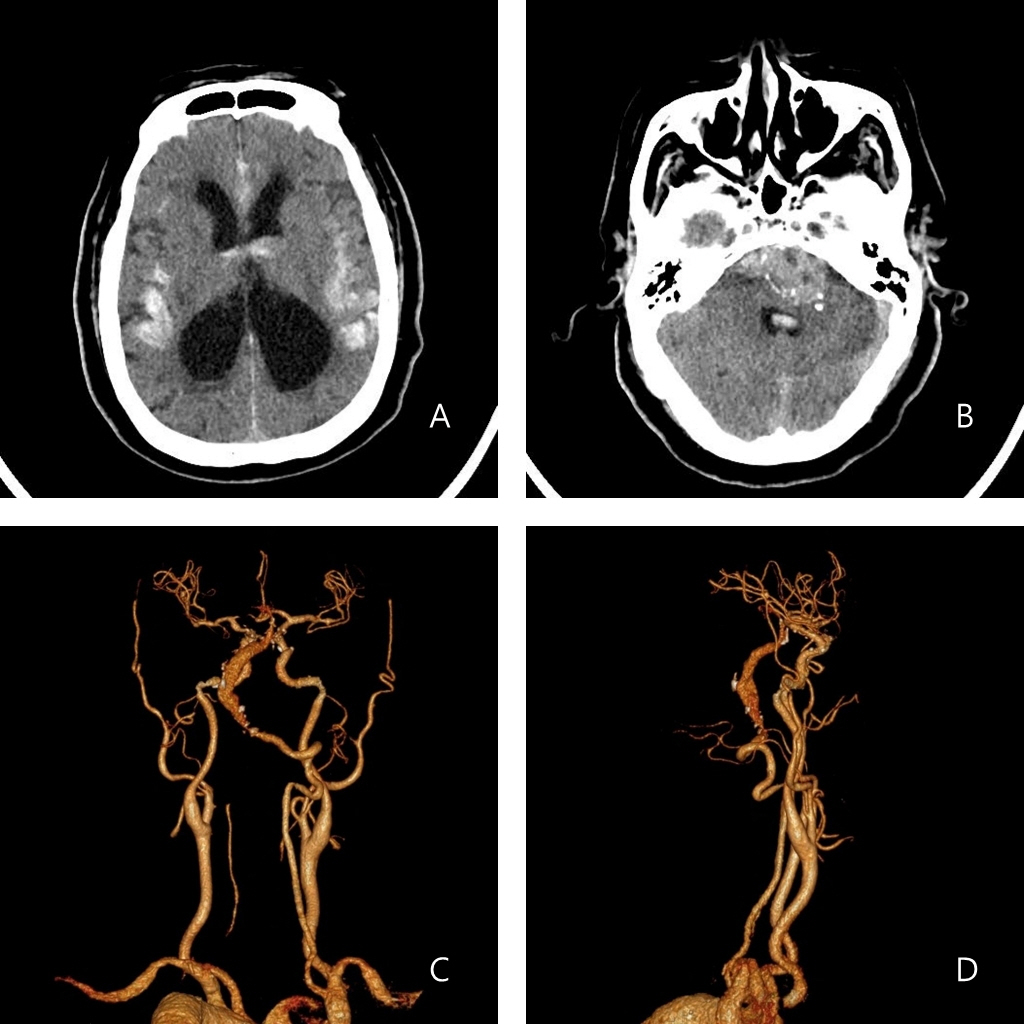
Fig. 1. (A) Computed tomography of the brain revealed subarachnoid hemorrhage with intraventricular hemorrhage and hydrocephalus. (B) Subarachnoid hemorrhage involved the posterior fossa near the fusiform aneurysm of the basilar artery, the basilar cistern, and both the sylvian cisterns. (C, D) Computed tomography angiography of the brain showing a giant vertebrobasilar dolichoectasia involving the right vestibular and basilar arteries.
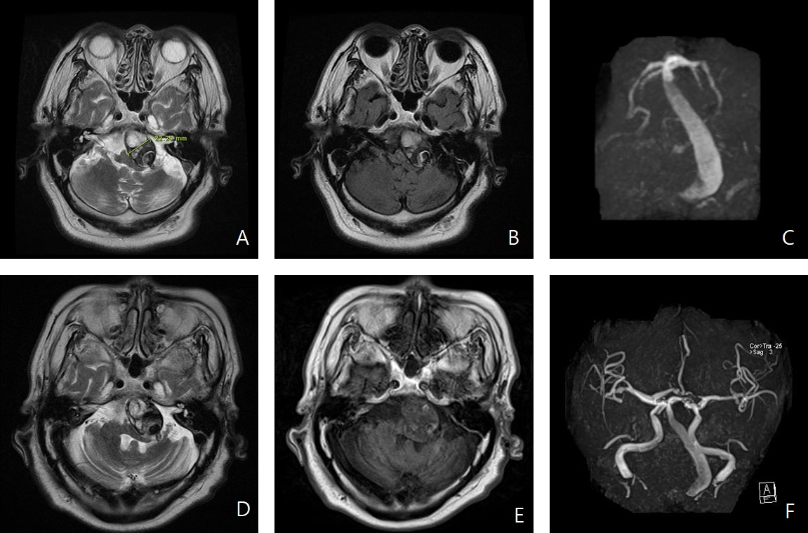
Fig. 2. (A, B) Magnetic resonance imaging (MRI) showing a giant vertebrobasilar dolichoectasia (22 mm in diameter) with a large mural thrombus within the tortuous basilar artery shifting the brainstem toward the right side. (C) Magnetic resonance angiography (MRA) demonstrating narrowing of the lumen of the basilar artery because of a thrombus. (D-F) MRI and MRA performed 5 months before the subarachnoid hemorrhage appeared showing no significant changes in the dimensions of the artery with vertebrobasilar dolichoectasia.
Reference
-
1. Del Brutto VJ, Ortiz JG, Biller J. Intracranial arterial dolichoectasia. Front Neurol. 2017; 8:344.
Article2. Pico F, Labreuche J, Amarenco P. Pathophysiology, presentation, prognosis, and management of intracranial arterial dolichoectasia. Lancet Neurol. 2015; 14:833–45.
Article3. Smoker WR, Price MJ, Keyes WD, Corbett JJ, Gentry LR. High-resolution computed tomography of the basilar artery: 1. normal size and position. AJNR Am J Neuroradiol. 1986; 7:55–60.4. Nakatomi H, Segawa H, Kurata A, Shiokawa Y, Nagata K, Kamiyama H, et al. Clinicopathological study of intracranial fusiform and dolichoectatic aneurysms: insight on the mechanism of growth. Stroke. 2000; 31:896–900.5. Mizutani T, Miki Y, Kojima H, Suzuki H. Proposed classification of nonatherosclerotic cerebral fusiform and dissecting aneurysms. Neurosurgery. 1999; 45:253–9.
Article6. Park JG, Kim BC, Lee SH, Choi SM, Park MS, Kim MK, et al. Brainstem infarction in patients with basilar artery dolichosis. J Korean Neurol Assoc. 2005; 23:318–23.7. Rahman EA, Trobe JD, Gebarski SS. Hemifacial spasm caused by vertebral artery dolichoectasia. Am J Ophthalmol. 2002; 133:854–6.
Article8. Ma X, Sun X, Yao J, Ni S, Gong J, Wang J, et al. Clinical analysis of trigeminal neuralgia caused by vertebrobasilar dolichoectasia. Neurosurg Rev. 2013; 36:573–7.
Article9. Siddiqui A, Chew NS, Miszkiel K. Vertebrobasilar dolichoectasia: a rare cause of obstructive hydrocephalus: case report. Br J Radiol. 2008; 81:e123–6.10. Sokolov AA, Husain S, Sztajzel R, Croquelois A, Lobrinus JA, Thaler D, et al. Fatal subarachnoid hemorrhage following ischemia in vertebrobasilar dolichoectasia. Medicine (Baltimore). 2016; 95:e4020.
Article11. Passero SG, Calchetti B, Bartalini S. Intracranial bleeding in patients with vertebrobasilar dolichoectasia. Stroke. 2005; 36:1421–5.
Article12. Saliou G, Sacho RH, Power S, Kostynskyy A, Willinsky RA, Tymianski M, et al. Natural history and management of basilar trunk artery aneurysms. Stroke. 2015; 46:948–53.
Article13. Coert BA, Chang SD, Do HM, Marks MP, Steinberg GK. Surgical and endovascular management of symptomatic posterior circulation fusiform aneurysms. J Neurosurg. 2007; 106:855–65.
Article14. Raphaeli G, Collignon L, De Witte O, Lubicz B. Endovascular treatment of posterior circulation fusiform aneurysms: single-center experience in 31 patients. Neurosurgery. 2011; 69:274–83.
Article
- Full Text Links
-
- Actions
-
Cited
- CITED
-
- Close
- Share
-
- Similar articles
-
- Rupture of the Giant Vertrbrobasilar Aneurysm After Hunterian Vertebral Artery Occlusion
- Lower Cranial Nerve Palsy Due to Vertebrobasilar Dolichoectasia
- Huge Vertebrobasilar Dolichoectasia
- Dolichoectasia of vertebrobasilar artery presenting as facial pain: a case report
- Treatment for subarachnoid hemorrhage due to ruptured posterior cerebral arterial dolichoectasia with aortic arch anomaly
