Concurrent Posterolateral Corner Injury Associated with a Schatzker Type 2 Tibial Plateau Fracture: A Case Report
- Affiliations
-
- 1Department of Orthopedic Surgery, Seoul Adventist Hospital, Sahmyook Medical Center, Seoul, Korea. tsnam74@gmail.com
- KMID: 2468671
- DOI: http://doi.org/10.12671/jkfs.2020.33.1.27
Abstract
- Isolated posterolateral corner (PLC) injury associated with a Schatzker type 2 fracture is a very rare combination of injuries. A male who was driving a motor vehicle was injured after a collision accident. The plain radiographs and computed tomography scans of the knee showed a Schatzker type 2 fracture of the tibial plateau, mostly in the anterolateral portion of tibial plateau, and an avulsion fragment on the fibular tip. Magnetic resonance imaging showed no injury to cruciate ligaments, medial collateral ligament, or any meniscal injury. We performed an open reduction operation and internal fixation for treating the fracture. Six months later, he complained of instability. At 11 months later after initial operation, we performed the second operation for stabilizing the PLC. We present here a rare case of an isolated PLC injury associated with a Schatzker type 2 fracture. We discuss the mechanism of injury and review similar cases.
Keyword
MeSH Terms
Figure
-

Fig. 1 The anteroposterior (A)/lateral (B) plain radiographs and coronal (C)/sagittal (D) computed tomography showed the lateral tibial plateau (LTP) fracture involving mainly the anterior portion of the LTP.
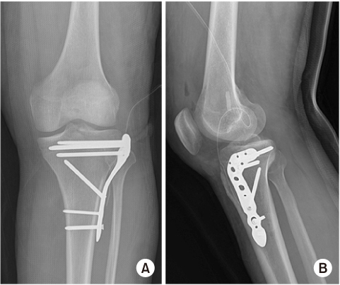
Fig. 2 The postoperative anteroposterior (A) and lateral (B) plain radiographs showed reduction of the lateral tibial plateau fracture.

Fig. 3 The coronal magnetic resonance imaging scans showed injury of the popliteus tendon (A; arrow) and biceps femoris avulsion (B; arrow).

Fig. 4 The postoperative anteroposterior (A) and lateral (B) plain radiographs showed reconstruction of the posterolateral corner.
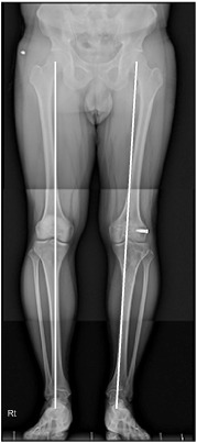
Fig. 5 Six months after the second operation, the whole standing anteroposterior plain radiograph showed neutral alignment (lines).
Reference
-
1. LaPrade RF, Wentorf FA, Fritts H, Gundry C, Hightower CD. A prospective magnetic resonance imaging study of the incidence of posterolateral and multiple ligament injuries in acute knee injuries presenting with a hemarthrosis. Arthroscopy. 2007; 23:1341–1347.
Article2. Yang BS, Bae WH, Ha JK, Lee DW, Jang HW, Kim JG. Posterolateral corner reconstruction using the single fibular sling method for posterolateral rotatory instability of the knee. Am J Sports Med. 2013; 41:1605–1612.
Article3. Terry GC, LaPrade RF. The posterolateral aspect of the knee. Anatomy and surgical approach. Am J Sports Med. 1996; 24:732–739.4. Seebacher JR, Inglis AE, Marshall JL, Warren RF. The structure of the posterolateral aspect of the knee. J Bone Joint Surg Am. 1982; 64:536–541.
Article5. LaPrade RF. Introduction and incidence of posterolateral knee injuries. In : LaPrade RF, editor. Posterolateral knee injuries: anatomy, evaluation, and treatment. New York: Thieme Medical Publishers;2006. p. 1–8.6. Stannard JP, Lopez R, Volgas D. Soft tissue injury of the knee after tibial plateau fractures. J Knee Surg. 2010; 23:187–192.
Article7. Zelle BA, Heaberlin JR, Murray MC. Posterolateral corner injury associated with a Schatzker type 2 tibial plateau fracture. Case Rep Orthop. 2015; 2015:527428.
Article8. Maheshwari J, Pandey VK, Mhaskar VA. Anterior tibial plateau fracture: an often missed injury. Indian J Orthop. 2014; 48:507–510.
Article9. Yoo JH, Lee JH, Chang CB. Pure varus injury to the knee joint. Clin Orthop Surg. 2015; 7:269–274.
Article
- Full Text Links
-
- Actions
-
Cited
- CITED
-
- Close
- Share
-
- Similar articles
-
- Analysis of Risk Factors for the Posterolateral Articular Depression and Status of Posterolateral Fragment in Lateral Condylar and Bicondylar Tibial Plateau Fractures with Joint Depression
- Evaluation of the Patterns of Fractures and the Soft Tissue Injury Using MRI in Tibial Plateau Fractures
- Prevalence of Meniscus Tear in Tibial Plateau Fractures
- Treatment of Tibial Condyle Fracture
- Operative Treatment of Tibial Plateau Fractures
