Perinatology.
2019 Dec;30(4):187-192. 10.14734/PN.2019.30.4.187.
Clinical Significance of Fetal Subcutaneous Thickness via Ultrasonography Biometry in the Third Trimester for Estimating Fetal Birth Weight
- Affiliations
-
- 1Division of Maternal and Fetal Medicine, Department of Obstetrics and Gynecology, Research Institute of Medical Science, Konkuk University School of Medicine, Seoul, Korea. hwanghs@kuh.ac.kr
- 2Department of Obstetrics and Gynecology, Ilsan Paik Hospital, Inje University College of Medicine, Goyang, Korea.
- KMID: 2468538
- DOI: http://doi.org/10.14734/PN.2019.30.4.187
Abstract
OBJECTIVE
In Korean population, information is lacking regarding fetal subcutaneous tissue thickness (SCTT) detected during pregnancy in the normal maternal condition. Thus, the aim of this study is to evaluate SCTT in the basic fetal biometry measurement plane, and to identify the clinical significance of SCTT in estimating fetal weight.
METHODS
In this retrospective observational study, 856 term pregnant women were recruited between 1st January 2013 and 31st December 2015. Estimated fetal weight (EFW) and fetal SCTT were measured routine ultrasonography within one week before delivery. The women were divided in two groups: SCTT group (n=46) and non-SCTT group (n=810). Pregnancy outcomes including birth weight (BW) and EFW were compared between the two groups.
RESULTS
The incidence of SCTT was 5.4% and no significant differences in parity, maternal age, maternal pre-pregnancy body mass index or gestational age at delivery were found between the groups. EFW, BW, amniotic fluid index, and cesarean section rate were higher in the SCTT group than in the non-SCTT group. The difference between EFW and BW was only significant in the SCTT group. Moreover, SCTT and EFW were positively correlated with BW (SCTT group: EFW 3,460±472 g vs. BW 3,779±496 g, P=0.013; non-SCTT group: EFW 3,011±436 g vs. BW 3,090±468 g, P=0.324).
CONCLUSION
Fetal SCTT detected during routine biometric ultrasonography evaluation in the third trimester of pregnancy could suggest larger BW than EFW. Therefore, physicians should pay careful attention in such cases during assessments for delivery.
Keyword
MeSH Terms
Figure
-
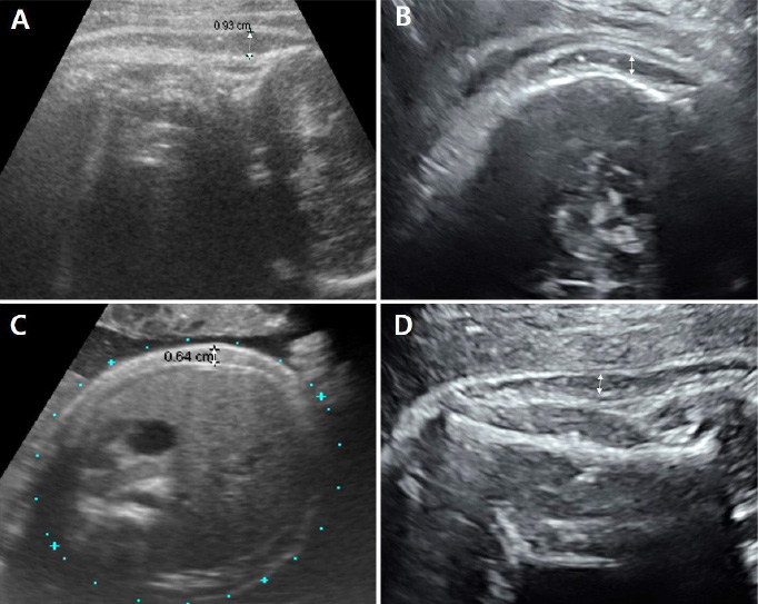
Fig. 1 Subcutaneous tissue thickness detected by ultrasonography in various planes (A, C: 37+2 weeks, male; B, D: 39+4 weeks, female). (A) Fetal ultrasonography image in the sagittal view at the posterior neck level, 9.3 mm. (B) Fetal ultrasonography image recorded in the plane used to determine humerus length, 3.2 mm. (C) Transverse view of the abdominal circumference, 6.4 mm. (D) Fetal ultrasonography image recorded in the plane used to determine head circumference, 3.6 mm.
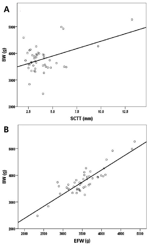
Fig. 2 Correlation between BW and EFW or SCTT. The correlation coefficient in each graph was (A) r=0.446, P=0.002 (B) r=0.875, P<0.001 by Spearman's rank correlation test. BW, birth weight; EFW, estimated fetal weight; SCTT, subcutaneous tissue thickness.
Reference
-
1. Warska A, Maliszewska A, Wnuk A, Szyszka B, Sawicki W, Cendrowski K. Current knowledge on the use of ultrasound measurements of fetal soft tissues for the assessment of pregnancy development. J Ultrason. 2018; 18:50–55.
Article2. Rotmensch S, Celentano C, Liberati M, Malinger G, Sadan O, Bellati U, et al. Screening efficacy of the subcutaneous tissue width/femur length ratio for fetal macrosomia in the non-diabetic pregnancy. Ultrasound Obstet Gynecol. 1999; 13:340–344.
Article3. Chauhan SP, West DJ, Scardo JA, Boyd JM, Joiner J, Hendrix NW. Antepartum detection of macrosomic fetus: clinical versus sonographic, including soft-tissue measurements. Obstet Gynecol. 2000; 95:639–642.
Article4. Luo H, Zhou S, Yang T, Liu S, Xu H. Ultrasonic multiple measurement estimation of fetal weight in parturient. Hua Xi Yi Ke Da Xue Xue Bao. 2001; 32:618–620.5. Lee W, Balasubramaniam M, Deter RL, Yeo L, Hassan SS, Gotsch F, et al. New fetal weight estimation models using fractional limb volume. Ultrasound Obstet Gynecol. 2009; 34:556–565.
Article6. Catalano PM, Tyzbir ED, Allen SR, McBean JH, McAuliffe TL. Evaluation of fetal growth by estimation of neonatal body composition. Obstet Gynecol. 1992; 79:46–50.7. Galan HL, Rigano S, Radaelli T, Cetin I, Bozzo M, Chyu J, et al. Reduction of subcutaneous mass, but not lean mass, in normal fetuses in Denver, Colorado. Am J Obstet Gynecol. 2001; 185:839–844.
Article8. Bernstein IM, Goran MI, Amini SB, Catalano PM. Differential growth of fetal tissues during the second half of pregnancy. Am J Obstet Gynecol. 1997; 176:28–32.
Article9. Maruotti GM, Saccone G, Martinelli P. Third trimester ultrasound soft-tissue measurements accurately predicts macrosomia. J Matern Fetal Neonatal Med. 2017; 30:972–976.
Article10. Larciprete G, Valensise H, Vasapollo B, Novelli GP, Parretti E, Altomare F, et al. Fetal subcutaneous tissue thickness (SCTT) in healthy and gestational diabetic pregnancies. Ultrasound Obstet Gynecol. 2003; 22:591–597.
Article11. Larciprete G, Valensise H, Barbati G, Di Pierro G, Jarvis S, Deaibess T, et al. Ultrasound-determined fetal subcutaneous tissue thickness for a birthweight prediction model. J Obstet Gynaecol Res. 2007; 33:635–640.
Article12. Hadlock FP, Deter RL, Harrist RB, Park SK. Estimating fetal age: computer-assisted analysis of multiple fetal growth parameters. Radiology. 1984; 152:497–501.
Article13. Chang TC, Robson SC, Boys RJ, Spencer JA. Prediction of the small for gestational age infant: which ultrasonic measurement is best? Obstet Gynecol. 1992; 80:1030–1038.14. Skovron ML, Berkowitz GS, Lapinski RH, Kim JM, Chitkara U. Evaluation of early third-trimester ultrasound screening for intrauterine growth retardation. J Ultrasound Med. 1991; 10:153–159.
Article15. Spady DW, Atrens MA, Szymanski WA. Effects of mother's smoking on their infants' body composition as determined by total body potassium. Pediatr Res. 1986; 20:716–719.
Article16. Forouzmehr A, Shahrokh A, Moulaei M. Estimation of birth weight using sonographically measured fetal abdominal subcutaneous tissue thickness. Iran J Radiol. 2004; 2:48–49.17. Chen L, Wu JJ, Chen XH, Cao L, Wu Y, Zhu LJ, et al. Measurement of fetal abdominal and subscapular subcutaneous tissue thickness during pregnancy to predict macrosomia: a pilot study. PLoS One. 2014; 9:e93077.
Article18. Abuelghar W, Khairy A, El Bishry G, Ellaithy M, Abd-Elhamid T. Fetal mid-thigh soft-tissue thickness: a novel method for fetal weight estimation. Arch Gynecol Obstet. 2014; 290:1101–1108.
Article19. Scioscia M, Scioscia F, Vimercati A, Caradonna F, Nardelli C, Pinto LR, et al. Estimation of fetal weight by measurement of fetal thigh soft-tissue thickness in the late third trimester. Ultrasound Obstet Gynecol. 2008; 31:314–320.
Article20. Kalantari M, Negahdari A, Roknsharifi S, Qorbani M. A new formula for estimating fetal weight: the impression of biparietal diameter, abdominal circumference, mid-thigh soft tissue thickness and femoral length on birth weight. Iran J Reprod Med. 2013; 11:933–938.21. Catalano PM, Thomas A, Huston-Presley L, Amini SB. Increased fetal adiposity: a very sensitive marker of abnormal in utero development. Am J Obstet Gynecol. 2003; 189:1698–1704.
Article22. Araujo Júnior E, Peixoto AB, Zamarian AC, Elito Júnior J, Tonni G. Macrosomia. Best Pract Res Clin Obstet Gynaecol. 2017; 38:83–96.
Article23. Tantanasis T, Daniilidis A, Giannoulis C, Tzafettas M, Dinas K, Loufopoulos A, et al. Sonographic assessment of fetal subcutaneous fat tissue thickness as an indicator of gestational diabetes. Eur J Obstet Gynecol Reprod Biol. 2010; 152:157–162.
Article
- Full Text Links
-
- Actions
-
Cited
- CITED
-
- Close
- Share
-
- Similar articles
-
- Clinical significance of sonographic soft markers: A review
- Estimation of Fetal Weight from Transabdominal Ultrasonic Measurements of Fetal Liver Length
- Measurements of Fetal Nuchal Skinfold Thickness in Normal Fetuses during Second Trimester
- Maternal Weight Gain Pattern and Birth Weight
- A Case of Fetal Cholelithiasis Related to Maternal Intrahepatic Cholestasis of Pregnancy
