Retinal Toxicity Following the Injection Ganciclovir into Silicone Oil-filled Eye to Treat Acute Retinal Necrosis
- Affiliations
-
- 1Department of Ophthalmology, Pusan National University School of Medicine, Yangsan, Korea. oph97@naver.com
- KMID: 2468484
- DOI: http://doi.org/10.3341/jkos.2020.61.1.111
Abstract
- PURPOSE
To report a case of retinal toxicity after an intravitreal ganciclovir injection to treat acute retinal necrosis in an eye filled with silicone oil.
CASE SUMMARY
A 56-year-old male presented with ocular pain and visual loss in his right eye. His best-corrected visual acuity was 20/25, inflammatory cells in the anterior chamber, multiple retinitis lesions and retinal vessel occlusions in the peripheral retina and vitreous opacity were showed. Acute retinal necrosis was suspected, anterior chamber polymerase chain reaction (PCR) test was done. Aciclovir 2,400 mg/day intravenously and ganciclovir 2.0 mg were administered by intravitreal injection. After 4 days, retinitis was worsened and PCR test was positive for varicella zoster virus. Ganciclovir intravitreal injections were increased twice a week. After 16 days, retinal detachment occurred, so scleral encircling, vitrectomy, laser photocoagulation, and silicone oil tamponade were conducted. Ganciclovir 1.0 mg was injected at the end of surgery. The patient's visual acuity decreased to hand motion, and multiple crystal deposits with multiple retinal hemorrhages were observed in the right eye the next day. Visual acuity did not recover and optical coherent tomography showed that the macula was thinned.
CONCLUSIONS
Visual loss seemed to be related with the retinal toxicity of ganciclovir. The increased local concentration due to the silicone oil tamponade is thought to have caused the toxicity.
MeSH Terms
-
Acyclovir
Anterior Chamber
Ganciclovir*
Hand
Herpesvirus 3, Human
Humans
Intravitreal Injections
Light Coagulation
Male
Middle Aged
Polymerase Chain Reaction
Retina
Retinal Detachment
Retinal Hemorrhage
Retinal Necrosis Syndrome, Acute*
Retinal Vessels
Retinaldehyde*
Retinitis
Silicon*
Silicones*
Visual Acuity
Vitrectomy
Acyclovir
Ganciclovir
Retinaldehyde
Silicon
Silicones
Figure
-
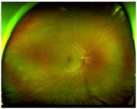
Figure 1 Ultra-widefield fundus image at the initial visit. Multiple retinal opaque was presented in the periphery. Mild vitreous haziness and retinal vasculitis were combined.
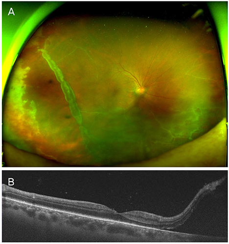
Figure 2 Ultra-widefield fundus image and optical coherent tomography at the sixteen day after (A) retinal detachment with giant retinal tear were noticed in the fundus examination. (B) Optical coherence tomography showed fovea center was not involved.
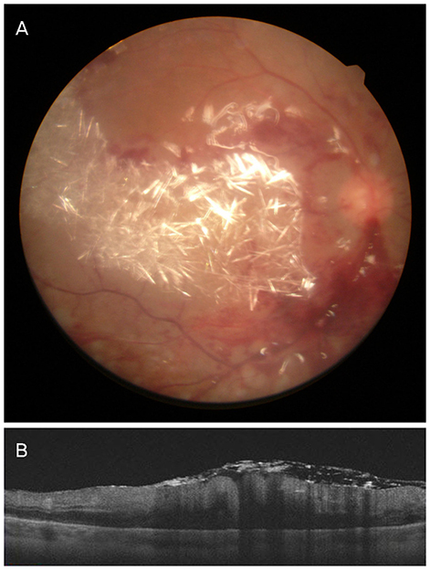
Figure 3 Fundus photo and optical coherent tomography at the next day of the surgery (A) multiple crystal deposit with multiple retinal hemorrhages was noticed on the fundus examination. (B) Optical coherence tomography showed severe macular edema.
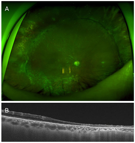
Figure 4 Ultra-widefield fundus image and optical coherent tomography at the sixth months after (A) there were no necrotic lesion, inflammation and retinal detachment on the fundus examination. (B) Optical coherence tomography image showed thinned macula.
Reference
-
1. Urayama A. Unilateral acute uveitis with relinal periarteritis and detachment. Jpn J Clin Ophthalmol. 1971; 25:607–619.2. Holland GN. Standard diagnostic criteria for the acute retinal necrosis syndrome. Executive Committee of the American Uveitis Society. Am J Ophthalmol. 1994; 117:663–667.3. Bernheim D, Germi R, Labetoulle M, et al. Time profile of viral DNA in aqueous humor samples of patients treated for varicella-zoster virus acute retinal necrosis by use of quantitative real-time PCR. J Clin Microbiol. 2013; 51:2160–2166.
Article4. Flaxel CJ, Yeh S, Lauer AK. Combination systemic and intravitreal antiviral therapy in the management of acute retinal necrosis syndrome (an American Ophthalmological Society thesis). Trans Am Ophthalmol Soc. 2013; 111:133–144.5. Saran BR, Maguire AM. Retinal toxicity of high dose intravitreal ganciclovir. Retina. 1994; 14:248–252.
Article6. Teoh SC, Ou X, Lim TH. Intravitreal ganciclovir maintenance injection for cytomegalovirus retinitis: efficacy of a low-volume, intermediate-dose regimen. Ophthalmology. 2012; 119:588–595.
Article7. Clarkson JG, Blumenkranz MS, Culbertson WW, et al. Retinal detachment following the acute retinal necrosis syndrome. Ophthalmology. 1984; 91:1665–1668.
Article8. Park SW, Shin MK, Byon IS, et al. Risk factors of retinal detachment after acute retinal necrosis. J Korean Ophthalmol Soc. 2013; 54:1694–1699.
Article9. Matsuo T. Vitrectomy and silicone oil tamponade as an initial surgery for retinal detachment after acute retinal necrosis syndrome. Ocul Immunol Inflamm. 2005; 13:91–94.10. Duker JS, Blumenkranz MS. Diagnosis and management of the acute retinal necrosis (ARN) syndrome. Surv Ophthalmol. 1991; 35:327–343.
Article11. Yang JW, Kim WJ, Park YH. Two cases of acute retinal necrosis treated with systemic antiviral drugs and intravitreal antiviral injections. J Korean Ophthalmol Soc. 2009; 50:794–799.
Article12. Hegazy HM, Kivilcim M, Peyman GA, et al. Evaluation of toxicity of intravitreal ceftazidime, vancomycin, and ganciclovir in a silicone oil-filled eye. Retina. 1999; 19:553–557.
Article13. Meshi A, Friehmann A, Sella S, et al. Intravitreal administration of antiviral agents in silicone oil-filled human eyes. Ophthalmol Retina. 2017; 1:288–293.
Article
- Full Text Links
-
- Actions
-
Cited
- CITED
-
- Close
- Share
-
- Similar articles
-
- Vitrectomy and Silicone Oil Tamponade to Prevent Retinal Detachment in Severe Acute Retinal Necrosis Syndrome
- The Management of Giant Retinal Tear by use of Perfluorodecalin(DK-line(R) and Silicone Oil
- Morphologic and Immunohistochemical Studies of Attached Retina in Intravitreal Silicone Oil
- Treatment of Acute Retinal Necrosis with Acute Kidney Injury after Intravenous Antiviral Injection
- A Clinical Study of Retinal Detachment Following Intraocular Silicone Oil Removal
