Pediatr Gastroenterol Hepatol Nutr.
2020 Jan;23(1):110-114. 10.5223/pghn.2020.23.1.110.
Gastric Xanthoma in the Pediatric Population: A Possible Herald for Malignancy?
- Affiliations
-
- 1Department of Undergraduate Studies, University of South Florida, Tampa, FL, USA.
- 2Department of Anesthesiology, Mount Sinai Medical Center, Miami, FL, USA.
- 3Department of Pediatric Gastroenterology, UT Southwestern Medical Center, Dallas, TX, USA.
- 4Department of Gastroenterology and Nutrition, Johns Hopkins All Children's Hospital, St. Petersburg, FL, USA. mwilsey1@jhmi.edu
- 5Department of Anatomic and Clinical Pathology, Johns Hopkins All Children's Hospital, St. Petersburg, FL, USA.
- KMID: 2467900
- DOI: http://doi.org/10.5223/pghn.2020.23.1.110
Abstract
- Gastric xanthoma is frequently an incidental finding on upper endoscopy in adults. Gastric xanthomas (GX) can be mistaken for malignancies and warrant prompt histologic diagnosis. The underlying etiology is not fully understood; however, it has been linked to Helicobacter pylori gastritis and gastric cancer. GX in the pediatric population is largely unreported in the literature. Because of the relative rarity, documentation with case reports are essential to provide as much data as possible to see if there is a correlation between GX and malignant potential in the pediatric population. Our group is reporting two cases, a 10-year-old male and a 7-year-old male, both who presented with chronic dysphagia, upper abdominal pain, nausea, vomiting, and loss of appetite. Upper endoscopies for both patients revealed small polypoid lesions located in the antrum with foamy histiocytes on histology, leading to the diagnosis of gastric xanthoma.
Keyword
MeSH Terms
Figure
-
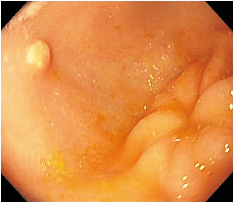
Fig. 1 Polypoid lesion near the gastric antrum as visible on upper endoscopy.
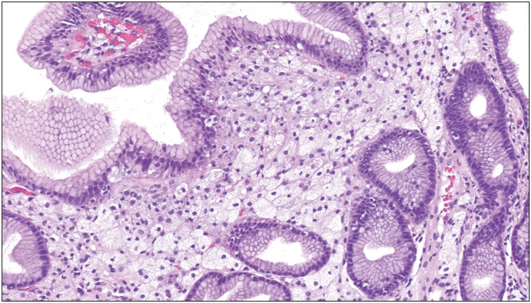
Fig. 2 The infiltrate in the lamina propria is composed of large foamy histiocytes with centrally-located, oval to round nuclei and open chromatin (H&E, ×200).
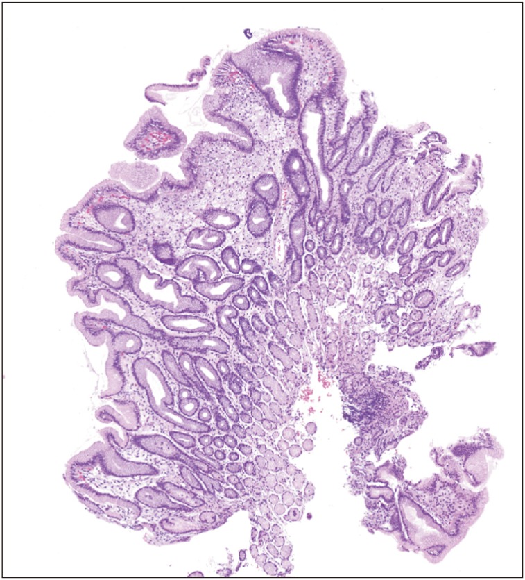
Fig. 3 Low power view shows a polypoid section of gastric mucosa with expansion of the lamina propria by numerous large cells with clear cytoplasm (H&E, ×40).
Reference
-
1. Basyigit S, Kefeli A, Asilturk Z, Sapmaz F, Aktas B. Gastric xanthoma: a review of the literature. Shiraz E Med J. 2015; 16.
Article2. Alzahrani M, Alqunaitir A, Alsohaibani M, Al-Rikabi AC. Gastric xanthelasma associated with hyperplastic polyp and mucosal erosions: report of an unusual case and literature review. Oxf Med Case Reports. 2018; 2018:omy051. PMID: 30151218.
Article3. Shibukawa N, Ouchi S, Wakamatsu S, Wakahara Y, Kaneko A. Gastric xanthoma is a predictive marker for metachronous and synchronous gastric cancer. World J Gastrointest Oncol. 2017; 9:327–332. PMID: 28868113.
Article4. Shibukawa N, Ouchi S, Wakamatsu S, Wakahara Y, Kaneko A. Gastric xanthoma is a predictive marker for early gastric cancer detected after helicobacter pylori eradication. Intern Med. 2019; 58:779–784. PMID: 30449773.5. Gasparetto M, Mara C, Graziella G, Gianmaria P, Francesca G. A rare case of pediatric gastric xanthoma: diagnosis and follow-up. J Gastroenterol Hepatol Res. 2013; 2:607–608.6. Halabi I, Yaseen M, Vesoulis Z, Nostrant TT. Multiple gastric xanthomas in a 3-year-old patient. Gastroenterol Hepatol. 2010; 6:181–184.
- Full Text Links
-
- Actions
-
Cited
- CITED
-
- Close
- Share
-
- Similar articles
-
- Gastric Xanthoma
- A Case of Type IV Hyperlipoproteinemia with Palmar Xanthoma, Tuberous Xanthoma, and Eruptive Xanthoma
- A Case of Gastric Xanthomatosis Scattered through Whole Gastric Mucosa
- A Case of Type IIb Hyperlipoproteinemia with Xanthoma Tuberosum, Xanthoma Planum and Xanthoma Striatum Palmare
- Two Cases of Diffuse Plane Xanthoma
