Tissue-Clearing Technique and Cutaneous Nerve Biopsies: Quantification of the Intraepidermal Nerve-Fiber Density Using Active Clarity Technique-Pressure Related Efficient and Stable Transfer of Macromolecules Into Organs
- Affiliations
-
- 1Department of Anatomy, Korea University College of Medicine, Seoul, Korea. irhyu@korea.ac.kr
- 2Department of Dermatology, Korea University College of Medicine, Seoul, Korea.
- 3Department of Neurology, Korea University College of Medicine, Seoul, Korea.
- 4Division of Brain Korea 21 Plus Program for Biomedical Science, Korea University College of Medicine, Seoul, Korea.
- KMID: 2467764
- DOI: http://doi.org/10.3988/jcn.2019.15.4.537
Abstract
- BACKGROUND AND PURPOSE
Cutaneous nerve biopsies based on two-dimensional analysis have been regarded as a creditable assessment tool for diagnosing peripheral neuropathies. However, advancements in methodological imaging are required for the analysis of intact structures of peripheral nerve fibers. A tissue-clearing and labeling technique facilitates three-dimensional imaging of internal structures in unsectioned, whole biological tissues without excessive time or labor costs. We sought to establish whether a tissue-clearing and labeling technique could be used for the diagnostic evaluation of peripheral neuropathies.
METHODS
Five healthy individuals and four patients with small-fiber neuropathy (SFN) and postherpetic neuralgia (PHN) were prospectively enrolled. The conventional methods of indirect immunofluorescence (IF) and bright-field immunohistochemistry (IHC) were adopted in addition to the tissue-clearing and labeling method called active clarity technique-pressure related efficient and stable transfer of macromolecules into organs (ACT-PRESTO) to quantify the intraepidermal nerve-fiber density (IENFD).
RESULTS
The mean IENFD values obtained by IF, bright-field IHC, and ACT-PRESTO in the healthy control group were 6.54, 6.44, and 90.19 fibers/mm², respectively; the corresponding values in the patients with SFN were 1.99, 2.32, and 48.12 fibers/mm², respectively, and 3.06, 2.87, and 47.21 fibers/mm², respectively, in the patients with PHN.
CONCLUSIONS
This study has shown that a tissue-clearing method provided not only rapid and highly reproducible three-dimensional images of cutaneous nerve fibers but also yielded reliable quantitative IENFD data. Quantification of the IENFD using a tissue-clearing and labeling technique is a promising way to improve conventional cutaneous nerve biopsies.
Keyword
MeSH Terms
Figure
-
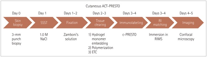
Fig. 1 Summary of the processing steps of the cutaneous ACT-PRESTO protocol, from separating the epidermis from biopsied skin samples to imaging with confocal microscopy. ACT-PRESTO: active clarity technique-pressure related efficient and stable transfer of macromolecules into organs, c-PRESTO: centrifugal-PRESTO, ETC: electrophoretic tissue clearing, RI: reflective index, RIMS: reflective-index matching solution, SSST: salt split skin test.
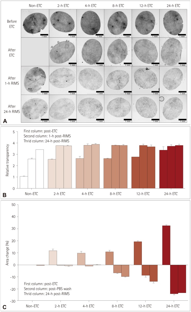
Fig. 2 Serial changes in transparency and area during the cutaneous ACT-PRESTO process, focusing on ETC and immersion in RIMS. A: Morphological changes in transparency according to the duration of ETC. B: Quantitative alterations of the relative transparency according to the duration of ETC. The improvement in relative transparency was proportional to the duration of ETC, but the differences were standardized after immersion in RIMS. C: Extent of area change related to the duration of ETC. The skin specimens consistently expanded after ETC in proportion to the duration of ETC. The enlarged tissues returned to their original size after washing with PBS. However, ETC durations exceeding 4 h resulted in irreversible shrinkage of the original tissues to an extent proportional to the ETC duration. ACT-PRESTO: active clarity technique-pressure related efficient and stable transfer of macromolecules into organs, ETC: electrophoretic tissue clearing, PBS: phosphate-buffered saline, RIMS: reflective-index matching solution.
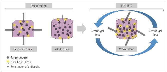
Fig. 3 Schematic diagram of immunohistochemistry for whole-tissue processing after tissue-clearing. The effectiveness of combinations of target antigens and specific antibodies was substantially impeded by the thicker whole tissue compared with conventional sectioned tissue. Tissues for c-PRESTO were centrifuged to obtain sufficient penetration depths of the primary and secondary antibodies, and so the technique could increase the probability of binding between target antigens and specific antibodies. c-PRESTO: centrifugal-pressure related efficient and stable transfer of macromolecules into organs.
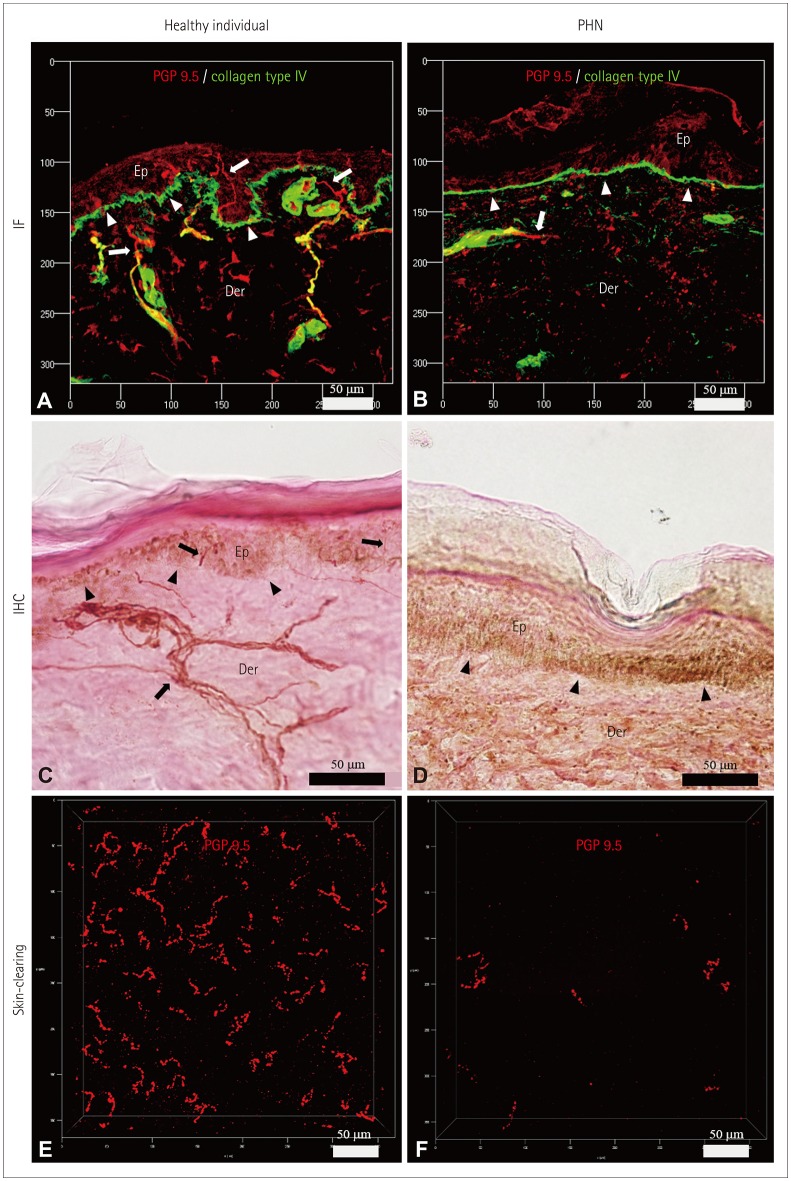
Fig. 4 Characteristics of the IENFs of a healthy participant (A, C, and E) and a PHN patient (B, D, and F). IENFs were quantified using the conventional methods of indirect IF (A and B) and IHC (C and D), as well as the ACT-PRESTO tissue-clearing technique (E and F). Cross-sectional views (A–D) and bird's-eye views (E and F) of IENFs. IF images (A and B) were obtained using confocal microscopy with a Plan-Apochromat 20×/0.8 M27 lens (maximum projection; stack size, 80 µm; stack step, 1 µm). IHC images (C and D) were produced using bright-field microscopy with a UPlanApo 40×/0.85 lens. Cutaneous nerve fibers (arrows) and the basal layer of the epidermis (arrowheads) (A–D). Images associated with skin-clearing (E and F) were also obtained using confocal microscopy with a Plan-Apochromat 20×/0.8 M27 lens (stack size, 200 µm; stack step, 1 µm). ACT-PRESTO: active clarity technique-pressure related efficient and stable transfer of macromolecules into organs, Der: dermis, Ep: epidermis, IENFs: intraepidermal nerve fibers, IF: immunofluorescence, IHC: immunohistochemistry, PGP 9.5: protein gene product 9.5, PHN: postherpetic neuralgia.
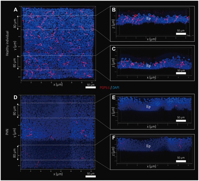
Fig. 5 Images of IENFs produced by the skin-clearing technique. IENFs of a healthy subject (A–C) and a PHN patient (D–F). Selected areas of three-dimensional images (A and D) in Fig. 4E and F, respectively, were optically sectioned at a thickness of 80 µm (B–F). All images were obtained and processed using confocal microscopy with a UPlanApo 40×/0.85 lens and the related software. Ep: epidermis, IENFs: intraepidermal nerve fibers, PGP 9.5: protein gene product 9.5, PHN: postherpetic neuralgia.
Reference
-
1. Lauria G, Cornblath DR, Johansson O, McArthur JC, Mellgren SI, Nolano M, et al. EFNS guidelines on the use of skin biopsy in the diagnosis of peripheral neuropathy. Eur J Neurol. 2005; 12:747–758. PMID: 16190912.
Article2. Joint Task Force of the EFNS and the PNS. European Federation of Neurological Societies/Peripheral Nerve Society Guideline on the use of skin biopsy in the diagnosis of small fiber neuropathy. Report of a joint task force of the European Federation of Neurological Societies and the Peripheral Nerve Society. J Peripher Nerv Syst. 2010; 15:79–92. PMID: 20626771.3. Devigili G, Tugnoli V, Penza P, Camozzi F, Lombardi R, Melli G, et al. The diagnostic criteria for small fibre neuropathy: from symptoms to neuropathology. Brain. 2008; 131:1912–1925. PMID: 18524793.
Article4. Mellgren SI, Nolano M, Sommer C. The cutaneous nerve biopsy: technical aspects, indications, and contribution. Handb Clin Neurol. 2013; 115:171–188. PMID: 23931780.5. Lauria G. Small fiber neuropathies. Curr Opin Neurol. 2005; 18:591–597. PMID: 16155446.6. Chung K, Wallace J, Kim SY, Kalyanasundaram S, Andalman AS, Davidson TJ, et al. Structural and molecular interrogation of intact biological systems. Nature. 2013; 497:332–337. PMID: 23575631.
Article7. Lee E, Choi J, Jo Y, Kim JY, Jang YJ, Lee HM, et al. ACT-PRESTO: rapid and consistent tissue clearing and labeling method for 3-dimensional (3D) imaging. Sci Rep. 2016; 6:18631. PMID: 26750588.
Article8. Gong H, Zeng S, Yan C, Lv X, Yang Z, Xu T, et al. Continuously tracing brain-wide long-distance axonal projections in mice at a one-micron voxel resolution. Neuroimage. 2013; 74:87–98. PMID: 23416252.
Article9. Li A, Gong H, Zhang B, Wang Q, Yan C, Wu J, et al. Micro-optical sectioning tomography to obtain a high-resolution atlas of the mouse brain. Science. 2010; 330:1404–1408. PMID: 21051596.
Article10. Kennedy WR, Wendelschafer-Crabb G. The innervation of human epidermis. J Neurol Sci. 1993; 115:184–190. PMID: 8482979.
Article11. McCarthy BG, Hsieh ST, Stocks A, Hauer P, Macko C, Cornblath DR, et al. Cutaneous innervation in sensory neuropathies: evaluation by skin biopsy. Neurology. 1995; 45:1848–1855. PMID: 7477980.
Article12. Gammon WR, Briggaman RA, Inman AO 3rd, Queen LL, Wheeler CE. Differentiating anti-lamina lucida and anti-sublamina densa anti-BMZ antibodies by indirect immunofluorescence on 1.0 M sodium chloride-separated skin. J Invest Dermatol. 1984; 82:139–144. PMID: 6363567.13. Rowbotham MC, Yosipovitch G, Connolly MK, Finlay D, Forde G, Fields HL. Cutaneous innervation density in the allodynic form of postherpetic neuralgia. Neurobiol Dis. 1996; 3:205–214. PMID: 8980021.
Article14. Oaklander AL, Romans K, Horasek S, Stocks A, Hauer P, Meyer RA. Unilateral postherpetic neuralgia is associated with bilateral sensory neuron damage. Ann Neurol. 1998; 44:789–795. PMID: 9818935.
Article15. Boucek P, Havrdova T, Voska L, Lodererova A, He L, Saudek F, et al. Epidermal innervation in type 1 diabetic patients: a 2.5-year prospective study after simultaneous pancreas/kidney transplantation. Diabetes Care. 2008; 31:1611–1612. PMID: 18443196.16. Schifitto G, Yiannoutsos C, Simpson DM, Adornato BT, Singer EJ, Hollander H, et al. Long-term treatment with recombinant nerve growth factor for HIV-associated sensory neuropathy. Neurology. 2001; 57:1313–1316. PMID: 11591856.
Article17. Helmchen F, Denk W. Deep tissue two-photon microscopy. Nat Methods. 2005; 12:932–940.
Article
- Full Text Links
-
- Actions
-
Cited
- CITED
-
- Close
- Share
-
- Similar articles
-
- Skin Biopsy: Emerging Method for Small Nerve Fiber Evaluation
- Skin biopsy: an emerging method for small nerve fiber evaluation
- Intercostal nerve transfer for the treatment of brachial plexus injury
- Assessment of Intraepidermal Nerve Fiber using Skin Biopsy in Diabetic Polyneuropathy
- Quantification of the Nerve Fiber of the Terminal Branches of the Typical Brachial Plexus
