Shear-Wave and Strain Ultrasound Elastography of the Supraspinatus and Infraspinatus Tendons in Patients with Idiopathic Adhesive Capsulitis of the Shoulder: A Prospective Case-Control Study
- Affiliations
-
- 1Department of Radiology, Kyung Hee University Hospital at Gangdong, Kyung Hee University School of Medicine, Seoul, Korea. jinooki@daum.net
- 2Department of Orthopaedic Surgery, Kyung Hee University Hospital at Gangdong, College of Medicine, Kyung Hee University, Seoul, Korea.
- 3Department of Radiology, Kyung Hee University Hospital, Seoul, Korea.
- 4Department of Radiology, Samsung Medical Center, Sungkyunkwan University School of Medicine, Seoul, Korea.
- 5Department of Radiology, Soonchunhyang Bucheon Hospital, Soonchunhyang University College of Medicine, Bucheon, Korea.
- 6Department of Medicine, Graduate School, Kyung Hee University, Seoul, Korea.
- KMID: 2467028
- DOI: http://doi.org/10.3348/kjr.2018.0918
Abstract
OBJECTIVE
To compare the elasticity of the supraspinatus tendon (SST) and infraspinatus tendon (IST) in patients with idiopathic adhesive capsulitis of the shoulder (ACS) with those in the control groups and to evaluate the relationship between age and tendon elasticity.
MATERIALS AND METHODS
The Institutional Review Board approved this prospective, case-control study, which was conducted between November 2017 and March 2018, and informed consent was obtained from all participants. Control groups comprised healthy individuals or those with asymptomatic contralateral shoulders. Twenty-five shoulders in 20 participants in the ACS group (14 women; 53.5 ± 7.9 years) and 24 shoulders in 18 participants in the control group (6 women; 52.6 ± 10.5 years) were included. Elastography was performed in the oblique coronal plane at the neutral shoulder position. Mean/maximum/minimum velocity and stiffness from the shear-wave ultrasound elastography (SWE) and strain ratio (subcutaneous fat/target-tendon) from the strain ultrasound elastography (SE) of the SST and IST were evaluated. Statistical analyses were performed using the Mann-Whitney U test, receiver operating characteristic (ROC) curve, and Spearman correlation.
RESULTS
Both velocity and stiffness in SWE were higher, and the strain ratio in SE was lower in participants with symptomatic shoulders than in those with normal shoulders (p < 0.001). SST- and IST-mean velocity, mean stiffness, and strain ratios showed excellent area under the ROC curve (> 0.970). The elastic modulus was little correlated with age (Ï = −0.340-0.239).
CONCLUSION
SWE and SE indicated that SST and IST were stiffer in patients with ACS than in those with normal shoulders regardless of aging.
MeSH Terms
Figure
-
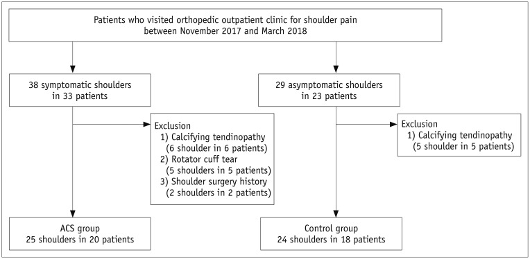
Fig. 1 Flow chart for patient selection.ACS = adhesive capsulitis of shoulder
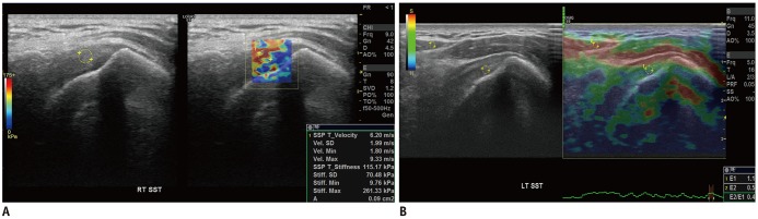
Fig. 2 Measurement of elastic modulus with shear-wave (A) and strain (B) ultrasound elastography.A. 65-year-old woman with ACS. Circular ROI of size 0.09-cm2 was set in target tendon at 0.5–1 cm from greater tuberosity (ROI1). Maximum, minimum, SD, and mean velocity in meter per second (m/s) and stiffness in kilopascal (kPa) are autogenerated. B. 75-year-old man with normal shoulder. Two circular ROIs were set in target tendon at 0.5–1 cm from greater tuberosity (ROI1) and subcutaneous fat (ROI2). Strain ratio (ROI2/ROI1) is autogenerated. ROI = region of interest, SD = standard deviation, SST = supraspinatus tendon
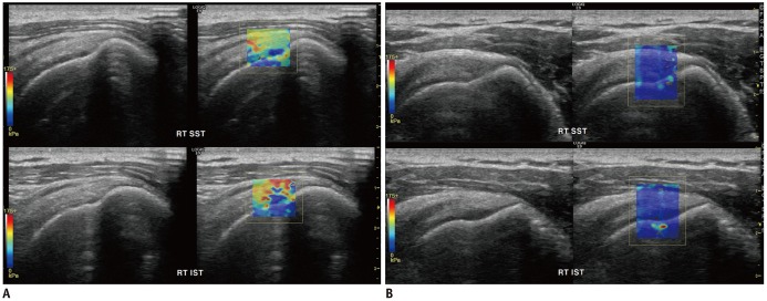
Fig. 3 Symptomatic shoulder in ACS group (A) and normal shoulder in control group (B) with shear-wave ultrasound elastography.A. 43-year-old man with ACS. Color in SST (upper) and IST (lower) is yellow to red, which indicates that tendons are stiff (SST-mean velocity, 4.12 m/s; SST-mean stiffness, 50.87 kPa; IST-mean velocity, 5.33 m/s; IST-mean stiffness, 85.33 kPa). B. 47-year-old man with normal shoulder. Color in SST (upper) and IST (lower) is entirely blue, which indicates that tendon is soft (SST-mean velocity, 1.06 m/s; SST-mean stiffness, 3.36 kPa; IST-mean velocity, 0.98 m/s; IST-mean stiffness, 2.88 kPa). IST = infraspinatus tendon
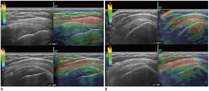
Fig. 4 Symptomatic shoulder in ACS group (A) and normal shoulder in control group (B) with strain ultrasound elastography.A. 51-year-old man with ACS. Color in SST (upper) and IST (lower) is green to dark blue, which indicates that tendons are stiff (SST-strain ratio, 0.06; IST-strain ratio, 0.05). B. 47-year-old man with normal shoulder. Color in box in SST (upper) and IST (lower) is yellowish, which indicates that tendon is soft (SST-strain ratio, 0.33; IST-strain ratio, 0.33).
Cited by 2 articles
-
Strain Ratio of Ultrasound Elastography for the Evaluation of Tendon Elasticity
Ke-Vin Chang, Wei-Ting Wu, Ing-Jeng Chen, Che-Yu Lin
Korean J Radiol. 2020;21(3):384-385. doi: 10.3348/kjr.2019.0737.Comparison of One- and Two-Region of Interest Strain Elastography Measurements in the Differential Diagnosis of Breast Masses
Hee Jeong Park, Sun Mi Kim, Bo La Yun, Mijung Jang, Bohyoung Kim, Soo Hyun Lee, Hye Shin Ahn
Korean J Radiol. 2020;21(4):431-441. doi: 10.3348/kjr.2019.0479.
Reference
-
1. Zuckerman JD, Rokito A. Frozen shoulder: a consensus definition. J Shoulder Elbow Surg. 2011; 20:322–325. PMID: 21051244.
Article2. Hsu JE, Anakwenze OA, Warrender WJ, Abboud JA. Current review of adhesive capsulitis. J Shoulder Elbow Surg. 2011; 20:502–514. PMID: 21167743.
Article3. Harris G, Bou-Haidar P, Harris C. Adhesive capsulitis: review of imaging and treatment. J Med Imaging Radiat Oncol. 2013; 57:633–643. PMID: 24283550.
Article4. Brue S, Valentin A, Forssblad M, Werner S, Mikkelsen C, Cerulli G. Idiopathic adhesive capsulitis of the shoulder: a review. Knee Surg Sports Traumatol Arthrosc. 2007; 15:1048–1054. PMID: 17333122.
Article5. Zappia M, Di Pietto F, Aliprandi A, Pozza S, De Petro P, Muda A, et al. Multi-modal imaging of adhesive capsulitis of the shoulder. Insights Imaging. 2016; 7:365–371. PMID: 27107871.
Article6. Homsi C, Bordalo-Rodrigues M, da Silva JJ, Stump XM. Ultrasound in adhesive capsulitis of the shoulder: is assessment of the coracohumeral ligament a valuable diagnostic tool? Skeletal Radiol. 2006; 35:673–678. PMID: 16724200.
Article7. Tandon A, Dewan S, Bhatt S, Jain AK, Kumari R. Sonography in diagnosis of adhesive capsulitis of the shoulder: a case-control study. J Ultrasound. 2017; 20:227–236. PMID: 28900523.
Article8. Wu CH, Chen WS, Wang TG. Elasticity of the coracohumeral ligament in patients with adhesive capsulitis of the shoulder. Radiology. 2016; 278:458–464. PMID: 26323030.
Article9. Michelin P, Delarue Y, Duparc F, Dacher JN. Thickening of the inferior glenohumeral capsule: an ultrasound sign for shoulder capsular contracture. Eur Radiol. 2013; 23:2802–2806. PMID: 23652851.
Article10. Klauser AS, Faschingbauer R, Jaschke WR. Is sonoelastography of value in assessing tendons? Semin Musculoskelet Radiol. 2010; 14:323–333. PMID: 20539957.
Article11. Ooi CC, Malliaras P, Schneider ME, Connell DA. “Soft, hard, or just right?” Applications and limitations of axial-strain sonoelastography and shear-wave elastography in the assessment of tendon injuries. Skeletal Radiol. 2014; 43:1–12. PMID: 23925561.
Article12. Pedersen M, Fredberg U, Langberg H. Sonoelastography as a diagnostic tool in the assessment of musculoskeletal alterations: a systematic review. Ultraschall Med. 2012; 33:441–446. PMID: 22744444.
Article13. DePalma AF. The classic. Loss of scapulohumeral motion (frozen shoulder). Ann Surg. 1952;135:193–204. Clin Orthop Relat Res. 2008; 466:552–560. PMID: 18264843.14. Crass JR, Craig EV, Feinberg SB. Clinical significance of sonographic findings in the abnormal but intact rotator cuff: a preliminary report. J Clin Ultrasound. 1988; 16:625–634. PMID: 3142923.
Article15. Fluss R, Faraggi D, Reiser B. Estimation of the Youden Index and its associated cutoff point. Biom J. 2005; 47:458–472. PMID: 16161804.
Article16. Zou KH, Tuncali K, Silverman SG. Correlation and simple linear regression. Radiology. 2003; 227:617–622. PMID: 12773666.
Article17. Landis JR, Koch GG. The measurement of observer agreement for categorical data. Biometrics. 1977; 33:159–174. PMID: 843571.
Article18. Tuite MJ, Small KM. Imaging evaluation of nonacute shoulder pain. AJR Am J Roentgenol. 2017; 209:525–533. PMID: 28537759.
Article19. Schmalzl J, Fenwick A, Boehm D, Gilbert F. The application of ultrasound elastography in the shoulder. J Shoulder Elbow Surg. 2017; 26:2236–2246. PMID: 29031414.
Article20. Taljanovic MS, Gimber LH, Becker GW, Latt LD, Klauser AS, Melville DM, et al. Shear-wave elastography: basic physics and musculoskeletal applications. Radiographics. 2017; 37:855–870. PMID: 28493799.
Article21. Ahn KS, Kang CH, Jeong WK. Contrast-enhanced ultrasonography in patients with adhesive capsulitis: preliminary experience. Iran J Radiol. 2017; 14:e33069.
Article22. Lee JC, Sykes C, Saifuddin A, Connell D. Adhesive capsulitis: sonographic changes in the rotator cuff interval with arthroscopic correlation. Skeletal Radiol. 2005; 34:522–527. PMID: 15999280.
Article23. Walmsley S, Osmotherly PG, Walker CJ, Rivett DA. Power Doppler ultrasonography in the early diagnosis of primary/idiopathic adhesive capsulitis: an exploratory study. J Manipulative Physiol Ther. 2013; 36:428–435. PMID: 23830711.
Article24. Li JQ, Tang KL, Wang J, Li QY, Xu HT, Yang HF, et al. MRI findings for frozen shoulder evaluation: is the thickness of the coracohumeral ligament a valuable diagnostic tool? PLoS One. 2011; 6:e28704. PMID: 22163326.
Article25. Greis C. Technology overview: SonoVue (Bracco, Milan). Eur Radiol. 2004; 14(Suppl 8):P11–P15. PMID: 15700328.26. Quaia E. Assessment of tissue perfusion by contrast-enhanced ultrasound. Eur Radiol. 2011; 21:604–615. PMID: 20927527.
Article27. Yasuda K, Hayashi K. Changes in biomechanical properties of tendons and ligaments from joint disuse. Osteoarthritis Cartilage. 1999; 7:122–129. PMID: 10367020.
Article28. Herbert RD, Crosbie J. Rest length and compliance of non-immobilised and immobilised rabbit soleus muscle and tendon. Eur J Appl Physiol Occup Physiol. 1997; 76:472–479. PMID: 9367288.
Article29. Ahn KS, Kang CH, Kim Y, Jeong WK. Diagnosis of adhesive capsulitis: comparison of contrast-enhanced MRI with noncontrast-enhanced MRI. Clin Imaging. 2015; 39:1061–1067. PMID: 26362354.
Article30. Connell D, Padmanabhan R, Buchbinder R. Adhesive capsulitis: role of MR imaging in differential diagnosis. Eur Radiol. 2002; 12:2100–2106. PMID: 12136330.
Article31. Gokalp G, Algin O, Yildirim N, Yazici Z. Adhesive capsulitis: contrast-enhanced shoulder MRI findings. J Med Imaging Radiat Oncol. 2011; 55:119–125. PMID: 21501399.
Article32. Gondim Teixeira PA, Balaj C, Chanson A, Lecocq S, Louis M, Blum A. Adhesive capsulitis of the shoulder: value of inferior glenohumeral ligament signal changes on T2-weighted fat-saturated images. AJR Am J Roentgenol. 2012; 198:W589–W596. PMID: 22623575.33. Lee SU, Joo SY, Kim SK, Lee SH, Park SR, Jeong C. Real-time sonoelastography in the diagnosis of rotator cuff tendinopathy. J Shoulder Elbow Surg. 2016; 25:723–729. PMID: 26794853.
Article34. Sasanuma H, Sugimoto H, Fujita A, Kanaya Y, Iijima Y, Saito T, et al. Characteristics of dynamic magnetic resonance imaging of idiopathic severe frozen shoulder. J Shoulder Elbow Surg. 2017; 26:e52–e57. PMID: 27539943.
Article35. Song KD, Kwon JW, Yoon YC, Choi SH. Indirect MR arthrographic findings of adhesive capsulitis. AJR Am J Roentgenol. 2011; 197:W1105–W1109. PMID: 22109326.
Article36. Yoon JP, Chung SW, Lee BJ, Kim HS, Yi JH, Lee HJ, et al. Correlations of magnetic resonance imaging findings with clinical symptom severity and prognosis of frozen shoulder. Knee Surg Sports Traumatol Arthrosc. 2017; 25:3242–3250. PMID: 26611904.
Article37. Hou SW, Merkle AN, Babb JS, McCabe R, Gyftopoulos S, Adler RS. Shear wave ultrasound elastographic evaluation of the rotator cuff tendon. J Ultrasound Med. 2017; 36:95–106. PMID: 27914201.
Article38. Kocyigit F, Kuyucu E, Kocyigit A, Herek DT, Savkin R, Aslan UB. Investigation of biomechanical characteristics of intact supraspinatus tendons in subacromial impingement syndrome: a cross-sectional study with real-time sonoelastography. Am J Phys Med Rehabil. 2016; 95:588–596. PMID: 26829089.39. Tudisco C, Bisicchia S, Stefanini M, Antonicoli M, Masala S, Simonetti G. Tendon quality in small unilateral supraspinatus tendon tears. Real-time sonoelastography correlates with clinical findings. Knee Surg Sports Traumatol Arthrosc. 2015; 23:393–398. PMID: 23771348.
Article40. Botar-Jid C, Damian L, Dudea SM, Vasilescu D, Rednic S, Badea R. The contribution of ultrasonography and sonoelastography in assessment of myositis. Med Ultrason. 2010; 12:120–126. PMID: 21173939.41. Baumer TG, Dischler J, Davis L, Labyed Y, Siegal DS, van Holsbeeck M, et al. Effects of age and pathology on shear wave speed of the human rotator cuff. J Orthop Res. 2018; 36:282–288. PMID: 28657192.
Article42. Hsiao MY, Chen YC, Lin CY, Chen WS, Wang TG. Reduced patellar tendon elasticity with aging: in vivo assessment by shear wave elastography. Ultrasound Med Biol. 2015; 41:2899–2905. PMID: 26304500.43. Arda K, Ciledag N, Aktas E, Aribas BK, Köse K. Quantitative assessment of normal soft-tissue elasticity using shear-wave ultrasound elastography. AJR Am J Roentgenol. 2011; 197:532–536. PMID: 21862792.
Article44. Slane LC, Martin J, DeWall R, Thelen D, Lee K. Quantitative ultrasound mapping of regional variations in shear wave speeds of the aging Achilles tendon. Eur Radiol. 2017; 27:474–482. PMID: 27236815.
Article45. Aubry S, Risson JR, Kastler A, Barbier-Brion B, Siliman G, Runge M, et al. Biomechanical properties of the calcaneal tendon in vivo assessed by transient shear wave elastography. Skeletal Radiol. 2013; 42:1143–1150. PMID: 23708047.
Article46. Peltz CD, Haladik JA, Divine G, Siegal D, van Holsbeeck M, Bey MJ. ShearWave elastography: repeatability for measurement of tendon stiffness. Skeletal Radiol. 2013; 42:1151–1156. PMID: 23640400.
Article47. Ewertsen C, Carlsen JF, Christiansen IR, Jensen JA, Nielsen MB. Evaluation of healthy muscle tissue by strain and shear wave elastography - Dependency on depth and ROI position in relation to underlying bone. Ultrasonics. 2016; 71:127–133. PMID: 27336792.
Article
