Image Quality and Radiation Dose in CT Venography Using Model-Based Iterative Reconstruction at 80 kVp versus Adaptive Statistical Iterative Reconstruction-V at 70 kVp
- Affiliations
-
- 1Department of Radiology, Pusan National University Yangsan Hospital, Yangsan, Korea. kschoo0618@naver.com
- 2Department of Radiology, Pusan National University Hospital, Busan, Korea.
- KMID: 2467027
- DOI: http://doi.org/10.3348/kjr.2018.0897
Abstract
OBJECTIVE
To compare the objective and subjective image quality indicators and radiation doses of computed tomography (CT) venography performed using model-based iterative reconstruction (MBIR) at 80 kVp and adaptive statistical iterative reconstruction (ASIR)-V at 70 kVp.
MATERIALS AND METHODS
Eighty-three patients who had undergone CT venography of the lower extremities with MBIR at 80 kVp (Group A; 21 men and 20 women; mean age, 55.5 years) or ASIR-V at 70 kVp (Group B; 18 men and 24 women; mean age, 57.3 years) were enrolled. Two radiologists retrospectively evaluated the objective (vascular enhancement, image noise, signal-to-noise ratio [SNR], contrast-to-noise ratio [CNR]) and subjective (quantum mottle, delineation of contour, venous enhancement) image quality indicators at the inferior vena cava and femoral and popliteal veins. Clinical information, radiation dose, reconstruction time, and objective and subjective image quality indicators were compared between groups A and B.
RESULTS
Vascular enhancement, SNR, and CNR were significantly greater in Group B than in Group A (p ≤ 0.015). Image noise was significantly lower in Group B (p ≤ 0.021), and all subjective image quality indicators, except for delineation of vein contours, were significantly better in Group B (p ≤ 0.021). Mean reconstruction time was significantly shorter in Group B than in Group A (1 min 43 s vs. 131 min 1 s; p < 0.001). Clinical information and radiation dose were not significantly different between the two groups.
CONCLUSION
CT venography using ASIR-V at 70 kVp was better than MBIR at 80 kVp in terms of image quality and reconstruction time at similar radiation doses.
MeSH Terms
Figure
-
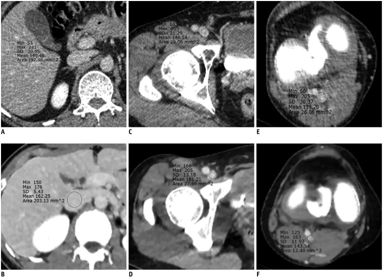
Fig. 1 Computed tomography images obtained using MBIR at 80 kVp (A, C, and E) or ASIR-V at 70 kVp (B, D, and F).Regions of interest were positioned within inferior vena cava (A, B) at level of left renal vein, within right femoral vein (C, D) at level of femoral head, and within right popliteal vein (E, F) at level of knee joint. Mean vascular enhancement (Hounsfield units) and subjective venous enhancement of three veins were greater with ASIR-V at 70 kVp (A, C, and E) than with MBIR at 80 kVp (B, D, and F). Quantum mottles of three veins were lower with ASIR-V at 70 kVp (grade 1, A; grade 2, C; and grade 2, E) than with MBIR at 80 kVp (grade 3, B; grade 4, D; and grade 3, F). ASIR = adaptive statistical iterative reconstruction, MBIR = model-based iterative reconstruction, SD = standard deviation
Reference
-
1. Raskob GE, Angchaisuksiri P, Blanco AN, Buller H, Gallus A, Hunt BJ, et al. ISTH Steering Committee for World Thrombosis Day. Thrombosis: a major contributor to global disease burden. Arterioscler Thromb Vasc Biol. 2014; 34:2363–2371. PMID: 25304324.
Article2. Huang W, Goldberg RJ, Anderson FA, Kiefe CI, Spencer FA. Secular trends in occurrence of acute venous thromboembolism: the Worcester VTE study (1985-2009). Am J Med. 2014; 127:829–839.e5. PMID: 24813864.
Article3. White RH. The epidemiology of venous thromboembolism. Circulation. 2003; 107(23 Suppl 1):I4–I8. PMID: 12814979.
Article4. Di Nisio M, van Es N, Büller HR. Deep vein thrombosis and pulmonary embolism. Lancet. 2016; 388:3060–3073. PMID: 27375038.
Article5. Bernardi E, Camporese G, Büller HR, Siragusa S, Imberti D, Berchio A, et al. Erasmus Study Group. Serial 2-point ultrasonography plus D-dimer vs whole-leg color-coded Doppler ultrasonography for diagnosing suspected symptomatic deep vein thrombosis: a randomized controlled trial. JAMA. 2008; 300:1653–1659. PMID: 18840838.6. Liu D, Peterson E, Dooner J, Baerlocher M, Zypchen L, Gagnon J, et al. Interdisciplinary Expert Panel on Iliofemoral Deep Vein Thrombosis (InterEPID). Diagnosis and management of iliofemoral deep vein thrombosis: clinical practice guideline. CMAJ. 2015; 187:1288–1296. PMID: 26416989.
Article7. Sasaki T, Fujimoto Y, Ishitoya S, Nabaa B, Watanabe N, Yamaki T, et al. Improved detectability of thromboses of the lower limb using low kilovoltage computed tomography. Medicine (Baltimore). 2018; 97:e9775. PMID: 29419670.
Article8. Jeong YJ, Choo KS, Nam KJ, Lee JW, Kim JY, Jung HJ, et al. Image quality and radiation dose of CT venography with double dose reduction using model based iterative reconstruction: comparison with conventional CT venography using filtered back projection. Acta Radiol. 2018; 59:546–552. PMID: 28766981.
Article9. Kim JH, Choo KS, Moon TY, Lee JW, Jeon UB, Kim TU, et al. Comparison of the image qualities of filtered back-projection, adaptive statistical iterative reconstruction, and model-based iterative reconstruction for CT venography at 80 kVp. Eur Radiol. 2016; 26:2055–2063. PMID: 26486938.
Article10. Cho ES, Chung JJ, Kim S, Kim JH, Yu JS, Yoon CS. CT venography for deep vein thrombosis using a low tube voltage (100 kVp) setting could increase venous enhancement and reduce the amount of administered iodine. Korean J Radiol. 2013; 14:183–193. PMID: 23482914.
Article11. Park CK, Choo KS, Jeon UB, Baik SK, Kim YW, Kim TU, et al. Image quality and radiation dose of 128-slice dual-source CT venography using low kilovoltage combined with high-pitch scanning and automatic tube current modulation. Int J Cardiovasc Imaging. 2013; 29(Suppl 1):47–51. PMID: 23748369.
Article12. Oda S, Utsunomiya D, Funama Y, Shimonobo T, Namimoto T, Itatani R, et al. Evaluation of deep vein thrombosis with reduced radiation and contrast material dose at computed tomography venography: clinical application of a combined iterative reconstruction and low-tube-voltage technique. Circ J. 2012; 76:2614–2622. PMID: 22784997.13. Kulkarni NM, Sahani DV, Desai GS, Kalva SP. Indirect computed tomography venography of the lower extremities using single-source dual-energy computed tomography: advantage of low-kiloelectron volt monochromatic images. J Vasc Interv Radiol. 2012; 23:879–886. PMID: 22633619.
Article14. Oda S, Utsunomiya D, Awai K, Takaoka H, Nakaura T, Katahira K, et al. Indirect computed tomography venography with a low-tube-voltage technique: reduction in the radiation and contrast material dose--a prospective randomized study. J Comput Assist Tomogr. 2011; 35:631–636. PMID: 21926861.15. Fujikawa A, Matsuoka S, Kuramochi K, Yoshikawa T, Yagihashi K, Kurihara Y, et al. Vascular enhancement and image quality of CT venography: comparison of standard and low kilovoltage settings. AJR Am J Roentgenol. 2011; 197:838–843. PMID: 21940570.
Article16. Matsuoka S, Hunsaker AR, Gill RR, Oliva IB, Trotman-Dickenson B, Jacobson FL, et al. Vascular enhancement and image quality of MDCT pulmonary angiography in 400 cases: comparison of standard and low kilovoltage settings. AJR Am J Roentgenol. 2009; 192:1651–1656. PMID: 19457830.
Article17. Pontone G, Muscogiuri G, Andreini D, Guaricci AI, Guglielmo M, Baggiano A, et al. Impact of a new adaptive statistical iterative reconstruction (ASIR)-V algorithm on image quality in coronary computed tomography angiography. Acad Radiol. 2018; 25:1305–1313. PMID: 29602723.
Article18. Goodenberger MH, Wagner-Bartak NA, Gupta S, Liu X, Yap RQ, Sun J, et al. Computed tomography image quality evaluation of a new iterative reconstruction algorithm in the abdomen (adaptive statistical iterative reconstruction-V) a comparison with model-based iterative reconstruction, adaptive statistical iterative reconstruction, and filtered back projection reconstructions. J Comput Assist Tomogr. 2018; 42:184–190. PMID: 28806318.
Article19. Kim HG, Lee HJ, Lee SK, Kim HJ, Kim MJ. Head CT: image quality improvement with ASIR-V using a reduced radiation dose protocol for children. Eur Radiol. 2017; 27:3609–3617. PMID: 28116512.
Article20. Lee S, Kwon H, Cho J. The detection of focal liver lesions using abdominal CT: a comparison of image quality between adaptive statistical iterative reconstruction V and adaptive statistical iterative reconstruction. Acad Radiol. 2016; 23:1532–1538. PMID: 27745816.
Article21. Kwon H, Cho J, Oh J, Kim D, Cho J, Kim S, et al. The adaptive statistical iterative reconstruction-V technique for radiation dose reduction in abdominal CT: comparison with the adaptive statistical iterative reconstruction technique. Br J Radiol. 2015; 88:20150463. PMID: 26234823.
Article22. Cham MD, Yankelevitz DF, Shaham D, Shah AA, Sherman L, Lewis A, et al. The Pulmonary Angiography-Indirect CT Venography Cooperative Group. Deep venous thrombosis: detection by using indirect CT venography. Radiology. 2000; 216:744–751. PMID: 10966705.
Article23. Landis JR, Koch GG. The measurement of observer agreement for categorical data. Biometrics. 1977; 33:159–174. PMID: 843571.
Article24. Sagara Y, Hara AK, Pavlicek W, Silva AC, Paden RG, Wu Q. Abdominal CT: comparison of low-dose CT with adaptive statistical iterative reconstruction and routine-dose CT with filtered back projection in 53 patients. AJR Am J Roentgenol. 2010; 195:713–719. PMID: 20729451.
Article25. Hussain FA, Mail N, Shamy AM, Suliman A, Saoudi A. A qualitative and quantitative analysis of radiation dose and image quality of computed tomography images using adaptive statistical iterative reconstruction. J Appl Clin Med Phys. 2016; 17:419–432. PMID: 27167261.
Article26. Brady SL, Moore BM, Yee BS, Kaufman RA. Pediatric CT: implementation of ASIR for substantial radiation dose reduction while maintaining pre-ASIR image noise. Radiology. 2014; 270:223–231. PMID: 23901128.
Article27. Kim HG, Chung YE, Lee YH, Choi JY, Park MS, Kim MJ, et al. Quantitative analysis of the effect of iterative reconstruction using a phantom: determining the appropriate blending percentage. Yonsei Med J. 2015; 56:253–261. PMID: 25510772.
Article28. Annoni AD, Andreini D, Pontone G, Formenti A, Petullà M, Consiglio E, et al. Ultra-low-dose CT for left atrium and pulmonary veins imaging using new model-based iterative reconstruction algorithm. Eur Heart J Cardiovasc Imaging. 2015; 16:1366–1373. PMID: 25911117.
Article29. Smith EA, Dillman JR, Goodsitt MM, Christodoulou EG, Keshavarzi N, Strouse PJ. Model-based iterative reconstruction: effect on patient radiation dose and image quality in pediatric body CT. Radiology. 2014; 270:526–534. PMID: 24091359.
Article30. Yasaka K, Katsura M, Hanaoka S, Sato J, Ohtomo K. High-resolution CT with new model-based iterative reconstruction with resolution preference algorithm in evaluations of lung nodules: comparison with conventional model-based iterative reconstruction and adaptive statistical iterative reconstruction. Eur J Radiol. 2016; 85:599–606. PMID: 26860673.
Article31. Deák Z, Grimm JM, Treitl M, Geyer LL, Linsenmaier U, Körner M, et al. Filtered back projection, adaptive statistical iterative reconstruction, and a model-based iterative reconstruction in abdominal CT: an experimental clinical study. Radiology. 2013; 266:197–206. PMID: 23169793.
Article32. Tang H, Yu N, Jia Y, Yu Y, Duan H, Han D, et al. Assessment of noise reduction potential and image quality improvement of a new generation adaptive statistical iterative reconstruction (ASIR-V) in chest CT. Br J Radiol. 2018; 91:20170521. PMID: 29076347.
Article33. Benz DC, Gräni C, Mikulicic F, Vontobel J, Fuchs TA, Possner M, et al. Adaptive statistical iterative reconstruction-V: impact on image quality in ultralow-dose coronary computed tomography angiography. J Comput Assist Tomogr. 2016; 40:958–963. PMID: 27560012.34. Waaijer A, Prokop M, Velthuis BK, Bakker CJ, de Kort GA, van Leeuwen MS. Circle of Willis at CT angiography: dose reduction and image quality--reducing tube voltage and increasing tube current settings. Radiology. 2007; 242:832–839. PMID: 17229873.
Article35. Huda W, Scalzetti EM, Levin G. Technique factors and image quality as functions of patient weight at abdominal CT. Radiology. 2000; 217:430–435. PMID: 11058640.
Article36. Kondratyev E, Karmazanovsky G. Low radiation dose 256-MDCT angiography of the carotid arteries: effect of hybrid iterative reconstruction technique on noise, artifacts, and image quality. Eur J Radiol. 2013; 82:2233–2239. PMID: 24094643.
Article37. Curry TS, Dowdey JE, Murry RC. Basic interactions between X-rays and matter. In : Curry TS, Dowdey JE, Murry RC, editors. Christensen's physics of diagnostic radiology. 4th ed. Philadelphia, PA: Lippincott Williams & Wilkins;1990. p. 61–69.38. Douketis JD, Crowther MA, Foster GA, Ginsberg JS. Does the location of thrombosis determine the risk of disease recurrence in patients with proximal deep vein thrombosis? Am J Med. 2001; 110:515–519. PMID: 11343664.
- Full Text Links
-
- Actions
-
Cited
- CITED
-
- Close
- Share
-
- Similar articles
-
- Comparison of Image Qualities of 80 kVp and 120 kVp CT Venography Using Model-Based Iterative Reconstruction at Same Radiation Dose
- Feasibility Study of Radiation Dose Reduction in Adult Female Pelvic CT Scan with Low Tube-Voltage and Adaptive Statistical Iterative Reconstruction
- Accuracy of Model-Based Iterative Reconstruction for CT Volumetry of Part-Solid Nodules and Solid Nodules in Comparison with Filtered Back Projection and Hybrid Iterative Reconstruction at Various Dose Settings: An Anthropomorphic Chest Phantom Study
- Combined Use of Automatic Tube Voltage Selection and Current Modulation with Iterative Reconstruction for CT Evaluation of Small Hypervascular Hepatocellular Carcinomas: Effect on Lesion Conspicuity and Image Quality
- Quantitative Analysis of the Effect of Iterative Reconstruction Using a Phantom: Determining the Appropriate Blending Percentage
