J Menopausal Med.
2019 Dec;25(3):164-171. 10.6118/jmm.19007.
Sonographic and Clinical Characteristics of Uterine Sarcoma Initially Misdiagnosed as Uterine Fibroid in Women in the Late Reproductive Age
- Affiliations
-
- 1Department of Obstetrics and Gynecology, Asan Medical Center, University of Ulsan College of Medicine, Seoul, Korea. kimsung@amc.seoul.kr
- 2Department of Obstetrics and Gynecology, Jeju National University School of Medicine, Jeju, Korea.
- KMID: 2466832
- DOI: http://doi.org/10.6118/jmm.19007
Abstract
OBJECTIVES
Uterine sarcoma is a rare malignant tumor, which is usually diagnosed in postmenopausal women. These sarcomas are occasionally misdiagnosed as uterine fibroids, thereby leading to delayed diagnosis in the advanced stages. We analyzed the sonographic and clinical characteristics of unexpected uterine sarcomas detected after surgery in women in the late reproductive age.
METHODS
The medical records of 61 patients preoperatively diagnosed with uterine leiomyomas through sonography but confirmed as uterine sarcomas after surgery from January 2005 to December 2018 at Asan Medical Center were retrospectively analyzed. We evaluated the clinical symptoms, sonographic findings, and Doppler indexes, and investigated whether there were any significant characteristics that could clearly differentiate uterine sarcoma from fibroids.
RESULTS
The most common clinical finding was increased mass size (15 patients, 24.6%), while 9 patients (14.8%) showed no symptoms. Ultrasonography showed that the maximum diameter of most fibroids was > 5 cm (49 patients, 80.3%), and the average diameter was 75.6 ± 36.3 mm. All the patients showed heterogeneous echogenicity in sonographic imaging. Secondary degeneration of the myomas was reported in 36 patients (59%), and approximately 90% (32/36, 88.9%) showed cystic changes. Of the 40 patients who underwent the evaluation of vascularity, 35 showed increased vascularity of the mass.
CONCLUSIONS
In this study, sarcomas misdiagnosed as leiomyomas were usually > 5 cm, and ultrasonography showed heterogeneous echogenicity and irregular cystic degeneration. No definite clinical symptoms were helpful; a thorough evaluation is necessary to rule out uterine sarcomas in women having uterine mass with these characteristics.
MeSH Terms
Figure
-
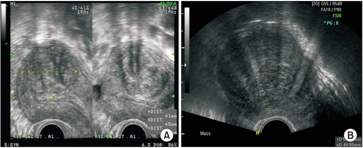
Fig. 1 (A) Internal shadowing of the mass and (B) fan-shaped shadowing.
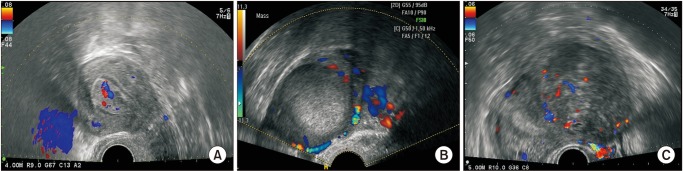
Fig. 2 (A) Vascularization on internal Doppler, (B) circumferential Doppler, and (C) both.
Reference
-
1. Major FJ, Blessing JA, Silverberg SG, Morrow CP, Creasman WT, Currie JL, et al. Prognostic factors in early-stage uterine sarcoma. A Gynecologic Oncology Group study. Cancer. 1993; 71(4 Suppl):1702–1709. PMID: 8381710.
Article2. Brooks SE, Zhan M, Cote T, Baquet CR. Surveillance, epidemiology, and end results analysis of 2677 cases of uterine sarcoma 1989–1999. Gynecol Oncol. 2004; 93:204–208. PMID: 15047237.
Article3. Aviram R, Ochshorn Y, Markovitch O, Fishman A, Cohen I, Altaras MM, et al. Uterine sarcomas versus leiomyomas: gray-scale and Doppler sonographic findings. J Clin Ultrasound. 2005; 33:10–13. PMID: 15690441.
Article4. Kurman RJ, Carcangiu ML, Herrington S, Young RH. WHO classification of tumours of female reproductive organs. 4th ed. Lyon: IARC;2014.5. Pritts EA, Vanness DJ, Berek JS, Parker W, Feinberg R, Feinberg J, et al. The prevalence of occult leiomyosarcoma at surgery for presumed uterine fibroids: a meta-analysis. Gynecol Surg. 2015; 12:165–177. PMID: 26283890.
Article6. Raine-Bennett T, Tucker LY, Zaritsky E, Littell RD, Palen T, Neugebauer R, et al. Occult uterine sarcoma and leiomyosarcoma: incidence of and survival associated With morcellation. Obstet Gynecol. 2016; 127:29–39. PMID: 26646120.7. Brohl AS, Li L, Andikyan V, Običan SG, Cioffi A, Hao K, et al. Age-stratified risk of unexpected uterine sarcoma following surgery for presumed benign leiomyoma. Oncologist. 2015; 20:433–439. PMID: 25765878.8. Chen I, Firth B, Hopkins L, Bougie O, Xie RH, Singh S. Clinical characteristics differentiating uterine sarcoma and fibroids. JSLS. 2018; 22:e2017.00066.
Article9. Ludovisi M, Moro F, Pasciuto T, Di Noi S, Giunchi S, Savelli L, et al. Imaging in gynecological disease (15): clinical and ultrasound characteristics of uterine sarcoma. Ultrasound Obstet Gynecol. 2019; 54:676–687. PMID: 30908820.10. Szabó I, Szánthó A, Csabay L, Csapó Z, Szirmai K, Papp Z. Color Doppler ultrasonography in the differentiation of uterine sarcomas from uterine leiomyomas. Eur J Gynaecol Oncol. 2002; 23:29–34. PMID: 11876388.11. Kurjak A, Kupesic S, Shalan H, Jukic S, Kosuta D, Ilijas M. Uterine sarcoma: a report of 10 cases studied by transvaginal color and pulsed Doppler sonography. Gynecol Oncol. 1995; 59:342–346. PMID: 8522252.
Article12. D'Angelo E, Prat J. Uterine sarcomas: a review. Gynecol Oncol. 2010; 116:131–139. PMID: 19853898.13. Mao J, Pfeifer S, Zheng XE, Schlegel P, Sedrakyan A. Population-based estimates of the prevalence of uterine sarcoma among patients with leiomyomata undergoing surgical treatment. JAMA Surg. 2015; 150:368–370. PMID: 25650751.
Article14. Zhao WC, Bi FF, Li D, Yang Q. Incidence and clinical characteristics of unexpected uterine sarcoma after hysterectomy and myomectomy for uterine fibroids: a retrospective study of 10,248 cases. Onco Targets Ther. 2015; 8:2943–2948. PMID: 26508879.15. Mbatani N, Olawaiye AB, Prat J. Uterine sarcomas. Int J Gynaecol Obstet. 2018; 143 Suppl 2:51–58. PMID: 30306577.
Article16. Van den Bosch T, Dueholm M, Leone FP, Valentin L, Rasmussen CK, Votino A, et al. Terms, definitions and measurements to describe sonographic features of myometrium and uterine masses: a consensus opinion from the Morphological Uterus Sonographic Assessment (MUSA) group. Ultrasound Obstet Gynecol. 2015; 46:284–298. PMID: 25652685.
Article
- Full Text Links
-
- Actions
-
Cited
- CITED
-
- Close
- Share
-
- Similar articles
-
- Uterine leiomyoma research
- Hemoperitoneum Caused by Spontaneous Rupture of Uterine Leiomyoma in a Perimenopausal Woman
- A Case of Uterine Fibroids Necrosis after Transarterial Embolization for Treatment of Uterine Fibroids
- The clinical responses of uterine artery embolization to treat uterine leiomyoma for 3 years
- A Case of Vaginal Expulsion of Submucosal Fibroid after Uterine Artery Embolization
