
Anat Cell Biol.
2019 Dec;52(4):478-485. 10.5115/acb.19.094.
Screening of the growth of thymus of human fetuses

- Affiliations
-
- 1Department of Anatomy, All India Institute of Medical Sciences Patna, Patna, India. dradilasghar2009@gmail.com
- 2Department of Basic Medical Sciences, College of Medicine, Majmaah University, Al-Majma'ah, Saudi Arabia.
- 3Department of Anaesthesiology, All India Institute of Medical Sciences Patna, Patna, India.
- 4Department of Anatomy, Uttar Pradesh University of Medical Sciences, Etawah, India.
- KMID: 2466702
- DOI: http://doi.org/10.5115/acb.19.094
Abstract
- The thymus is a lymphoepithelial organ, and its morphometry is commonly utilized for surveillance of the immunological status of fetus and neonates. Many studies showed that fetal thymus size is used as a prognostic indicator for pregnancy-related disorders such as eclampsia, preterm labor, and gestational diabetes. The study aims to establish reference ranges of the normal fetal thymus size between 12 and 40 weeks of gestational age (GA). The study was conducted on 89 fetuses. They were dissected to capture the morphometry of thymus: transverse diameter, perimeter, and weight. Considering these parameters were dependent variables of GA and gestational weight (GW). Their relationship was studied by a multiple regression model. The best fit models in predicting thymic dimensions as a function of GA and GW were determined using regression analysis. Mean transverse diameter, perimeter, and thymus weight was 33.45±2.91 mm, 125.72±55.4 mm, and 3.078±3.06 g, respectively. They were increased throughout pregnancy as GA and GW advanced. The regression equation for a transverse diameter of the thymus as a function of GA was (0.303×GA-4.885, R²=0.8196) and for the perimeter of the thymus was (1.0212×GA-15.24, R²=0.8666). Reference ranges and baseline data of the normal fetal thymic dimensions between 12 and 40 weeks of GA have been established.
MeSH Terms
Figure
-
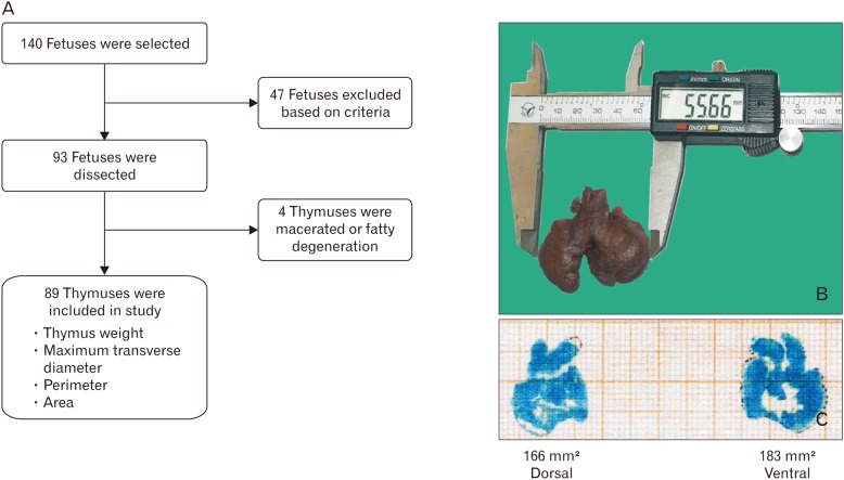
Fig. 1 (A) Methodology for study of growth pattern of thymus. (B) Measurement of Transverse diameter in dissected thymus by Digital Vernier Caliper. (C) Measurement of perimeter and area by taking impression on graph paper.
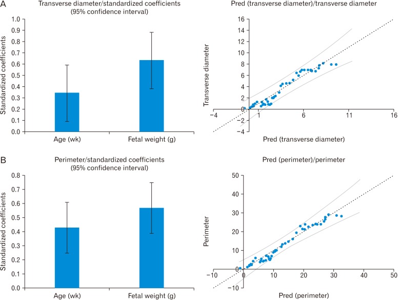
Fig. 2 Multiple regression analysis of transverse diameter with gestational age and weight (A) and perimeter with gestational age and weight (B).
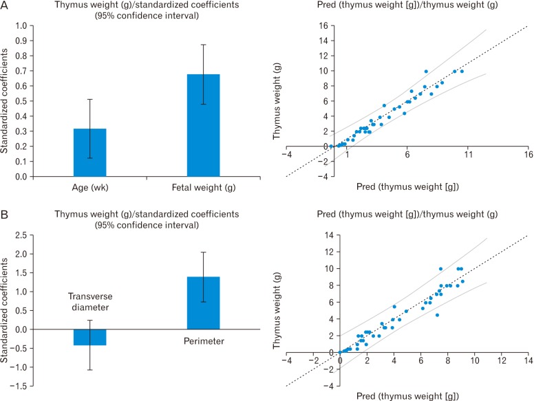
Fig. 3 Multiple regression analysis of thymus weight with gestational age and weight (A) and thymus weight with transverse diameter and perimeter of thymus (B).
Reference
-
1. Man J, Hutchinson JC, Ashworth M, Jeffrey I, Heazell AE, Sebire NJ. Organ weights and ratios for postmortem identification of fetal growth restriction: utility and confounding factors. Ultrasound Obstet Gynecol. 2016; 48:585–590. PMID: 27781326.2. Ciofani M, Zúñiga-Pflücker JC. The thymus as an inductive site for T lymphopoiesis. Annu Rev Cell Dev Biol. 2007; 23:463–493. PMID: 17506693.3. Jeppesen DL, Hasselbalch H, Nielsen SD, Sørensen TU, Ersbøll AK, Valerius NH, Heilmann C. Thymic size in preterm neonates: a sonographic study. Acta Paediatr. 2003; 92:817–822. PMID: 12892161.4. Toti P, De Felice C, Stumpo M, Schürfeld K, Di Leo L, Vatti R, Bianciardi G, Buonocore G, Seemayer TA, Luzi P. Acute thymic involution in fetuses and neonates with chorioamnionitis. Hum Pathol. 2000; 31:1121–1128. PMID: 11014581.5. Pagenkemper M, Diemert A. Monitoring fetal immune development in human pregnancies: current concepts and future goals. J Reprod Immunol. 2014; 104-105:49–53. PMID: 25124491.6. Di Naro E, Cromi A, Ghezzi F, Raio L, Uccella S, D'Addario V, Loverro G. Fetal thymic involution: a sonographic marker of the fetal inflammatory response syndrome. Am J Obstet Gynecol. 2006; 194:153–159. PMID: 16389025.7. Sakaguchi S. The origin of FOXP3-expressing CD4+ regulatory T cells: thymus or periphery. J Clin Invest. 2003; 112:1310–1312. PMID: 14597756.8. Cox P, Marton T. Pathological assessment of intrauterine growth restriction. Best Pract Res Clin Obstet Gynaecol. 2009; 23:751–764. PMID: 19854107.9. Yinon Y, Zalel Y, Weisz B, Mazaki-Tovi S, Sivan E, Schiff E, Achiron R. Fetal thymus size as a predictor of chorioamnionitis in women with preterm premature rupture of membranes. Ultrasound Obstet Gynecol. 2007; 29:639–643. PMID: 17471450.10. Cromi A, Ghezzi F, Raffaelli R, Bergamini V, Siesto G, Bolis P. Ultrasonographic measurement of thymus size in IUGR fetuses: a marker of the fetal immunoendocrine response to malnutrition. Ultrasound Obstet Gynecol. 2009; 33:421–426. PMID: 19306477.11. Mohamed N, Eviston DP, Quinton AE, Benzie RJ, Kirby AC, Peek MJ, Nanan RK. Smaller fetal thymuses in pre-eclampsia: a prospective cross-sectional study. Ultrasound Obstet Gynecol. 2011; 37:410–415. PMID: 21308839.12. Olearo E, Oberto M, Oggè G, Botta G, Pace C, Gaglioti P, Todros T. Thymic volume in healthy, small for gestational age and growth restricted fetuses. Prenat Diagn. 2012; 32:662–667. PMID: 22544629.13. Aaby P, Marx C, Trautner S, Rudaa D, Hasselbalch H, Jensen H, Lisse I. Thymus size at birth is associated with infant mortality: a community study from Guinea-Bissau. Acta Paediatr. 2002; 91:698–703. PMID: 12162605.14. Park HY, Hertz-Picciotto I, Petrik J, Palkovicova L, Kocan A, Trnovec T. Prenatal PCB exposure and thymus size at birth in neonates in Eastern Slovakia. Environ Health Perspect. 2008; 116:104–109. PMID: 18197307.15. Varga I, Uhrinova A, Toth F, Mistinova J. Assessment of the thymic morphometry using ultrasound in full-term newborns. Surg Radiol Anat. 2011; 33:689–695. PMID: 21442251.16. Acharya P, Acharya A. Evaluation of applicability of standard growth curves to Indian women by fetal biometry. J S Asian Fed Obstet Gynecol. 2009; 1:55–61.17. Muñoz-Chápuli M, Gámez F, Bravo C, Ortiz L, Pérez R, De León-Luis JA. The thy-box for sonographic assessment of the fetal thymus: nomogram and review of the literature. J Ultrasound Med. 2015; 34:853–858. PMID: 25911720.18. Cho JY, Min JY, Lee YH, McCrindle B, Hornberger LK, Yoo SJ. Diameter of the normal fetal thymus on ultrasound. Ultrasound Obstet Gynecol. 2007; 29:634–638. PMID: 17385216.19. Murao F, Takamori H, Hata K, Hata T, Kitao M. Fetal liver measurements by ultrasonography. Int J Gynaecol Obstet. 1987; 25:381–385. PMID: 2889632.20. Ho SS, Metreweli C. Normal fetal thyroid volume. Ultrasound Obstet Gynecol. 1998; 11:118–122. PMID: 9549838.21. Gloor JM, Breckle RJ, Gehrking WC, Rosenquist RG, Mulholland TA, Bergstralh EJ, Ramin KD, Ogburn PL Jr. Fetal renal growth evaluated by prenatal ultrasound examination. Mayo Clin Proc. 1997; 72:124–129. PMID: 9033544.22. Zalel Y, Perlitz Y, Gamzu R, Peleg D, Ben-Ami M. In-utero development of the fetal colon and rectum: sonographic evaluation. Ultrasound Obstet Gynecol. 2003; 21:161–164. PMID: 12601839.23. Gur EB, Gur MS, Ince O, Kasap E, Genc M, Tatar S, Bugday S, Turan GA, Guclu S. Vitamin D deficiency in pregnancy may affect fetal thymus development. Ginekol Pol. 2016; 87:378–383. PMID: 27304655.24. Gamez F, De Leon-Luis J, Pintado P, Perez R, Robinson JN, Antolin E, Ortiz-Quintana L, Santolaya-Forgas J. Fetal thymus size in uncomplicated twin and singleton pregnancies. Ultrasound Obstet Gynecol. 2010; 36:302–307. PMID: 20131331.25. Tangshewinsirikul C, Panburana P. Sonographic measurement of fetal thymus size in uncomplicated singleton pregnancies. J Clin Ultrasound. 2017; 45:150–159. PMID: 27862004.26. Musilova I, Hornychova H, Kostal M, Jacobsson B, Kacerovsky M. Ultrasound measurement of the transverse diameter of the fetal thymus in pregnancies complicated by the preterm prelabor rupture of membranes. J Clin Ultrasound. 2013; 41:283–289. PMID: 23505029.27. Hansen K, Sung CJ, Huang C, Pinar H, Singer DB, Oyer CE. Reference values for second trimester fetal and neonatal organ weights and measurements. Pediatr Dev Pathol. 2003; 6:160–167. PMID: 12548377.28. Guihard-Costa AM, Ménez F, Delezoide AL. Organ weights in human fetuses after formalin fixation: standards by gestational age and body weight. Pediatr Dev Pathol. 2002; 5:559–578. PMID: 12399830.
- Full Text Links
-
- Actions
-
Cited
- CITED
-
- Close
- Share
-
- Similar articles
-
- Weight and morphologic development of prenatal human thymus
- Renal Artery Pulsatility Index and Renal Volume: Normal Fetuses versus Growth-Retarded Fetuses
- Approximate Entropy: Analysis of Fetal Heart Rate Variability in Normal and Growth Retarded Fetuses
- The Distribution of MIC2 Antigen (CD99) Expression on Various Mucosa-Associated Lymphoid Tissue of Human Embryos and Fetuses
- Expression Pattern of Immunoproteasome Subunits in Human Thymus


