Korean Lepr Bull.
2019 Dec;52(1):55-61. 10.33161/klb.2019.52.1.55.
Relapsed Leprosy with Multiple Ulcerative Skin Lesions: A Case Report
- Affiliations
-
- 1Department of Dermatology, Samsung Changwon Hospital, Sungkyunkwan University School of Medicine, Korea. rhwodhks83@naver.com
- 2Institute for Leprosy Research, Korean Hansen Welfare Association, Korea.
- KMID: 2466493
- DOI: http://doi.org/10.33161/klb.2019.52.1.55
Abstract
- Leprosy is a chronic infectious and granulomatous disease caused by Mycobacterium leprae. It is treated with a multidrug therapy (MDT), which is consisted of dapsone, rifampicin, and clofazimine. However, there were relapsed leprosy associated with various predisposing factors; persisting organism, multiple involved skin lesions and nerves, HIV infection, monotherapy, inadequate and irregular therapy. Early or late relapses were observed in leprosy. Early onset relapses may occur due to insufficient treatment, and late relapses are probably with persistent bacilli and drug resistant organisms. Herein, we report on an interesting case of a 78-year-old man presented with relapsed leprosy associated with ulcerative skin lesions. The patient was diagnosed with lepromatous leprosy about 40 years ago, and he was treated with dapsone monotherapy and MDT. Our case is thought to have occurred due to persistent bacilli related to irregular therapy.
MeSH Terms
Figure
-
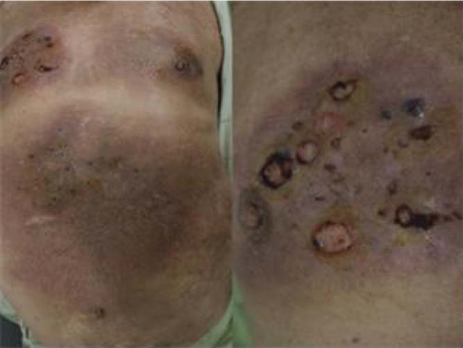
Fig. 1 (A) Erythematosus plaques with ulcerative skin lesions on the trunk (B) Multiple painful ulcers on the chest

Fig. 2 Histopathologic examination shows diffuse inflammatory cell infiltration in dermis (A) and subcutis (B) separated from subepidermal grenz zone (H&E, ×40), and also positive staining with acid-fast bacilli stain (×400) (C)
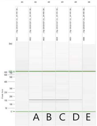
Fig. 3 PCR results show positive band on 157 bp. Lane (A) sample of tissue; (B) sample of tissue; (C) sample of paraffine; (D) positive control; (E) negative control.
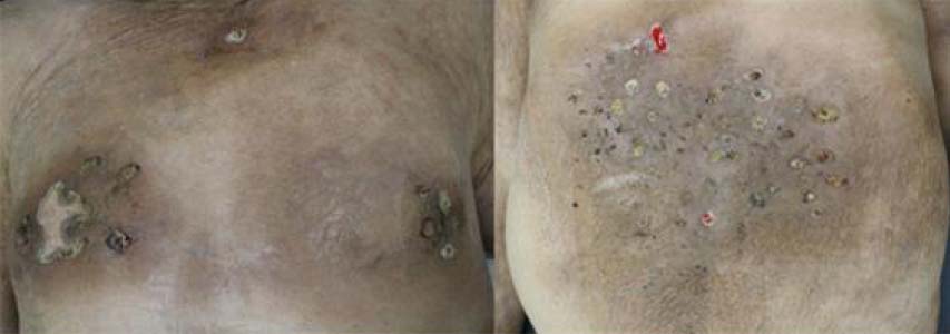
Fig. 4 After treatments, skin lesions on chest (A) and lower back (B) were showed scarring changes with crusts.
Reference
-
1. Eichelmann K, González González SE, Salas-Alanis JC, Ocampo-Candiani J. Leprosy. An update: definition, pathogenesis, classification, diagnosis, and treatment. Actas Dermosifiliogr. 2013; 104:554–556.
Article2. Lastória JC, Abreu MA. Leprosy: review of the epidemiological, clinical, and etiopathogenic aspects - part 1. An Bras Dermatol. 2014; 89:205–218.
Article3. Noordeen SK. Elimination of leprosy as a public health problem: progress and prospects. Bull World Health Organ. 1995; 73:1–6.4. Pattyn SR, Groenen G, Bourland J, De Muynck A, Grillone S, Grossetete G, et al. The incubation time of relapses after treatment of multibacillary leprosy with rifampicin containing regimens. Eur J Epidemiol. 1988; 4:231–234.
Article5. Park JM, Kim JP, Kang KH. New patient of Hansen's disease in young Korean man. Korean Lepr Bull. 2018; 51:23–28.
Article6. Han XY, Quintanilla M. Diffuse Lepromatous Leprosy Due to Mycobacterium lepromatosis in Quintana Roo, Mexico. J Clin Microbiol. 2015; 53:3695–3698.
Article7. Kaimal S, Thappa DM. Relapse in leprosy. Indian J Dermatol Venereol Leprol. 2009; 75:126–135.
Article8. The Leprosy Unit, WHO. Risk of relapse in leprosy. Indian J Lepr. 1995; 67:13–26.9. Becx-Bleumink M. Relapses among leprosy patients treated with multidrug therapy: experience in the leprosy control program of the All Africa Leprosy and Rehabilitation Training Center (ALERT) in Ethiopia; practicaldifficulties with diagnosing relapses; operational procedures and criteria for diagnosing relapses. Int J Lepr Other Mycobact Dis. 1992; 60:421–435.10. Kim JP. The summary of recent relapse leprosy cases in Korea (2003~2007). Korean Lepr Bull. 2008; 41:27–35.11. Ramu G. Clinical features and diagnosis of relapses in leprosy. Indian J Lepr. 1995; 67:45–59.12. Shetty VP, Wakade AV, Ghate SD, Pai VV, Ganapati RR, Antia NH. Clinical, histopathological and bacteriological study of 52 referral MB cases relapsing after MDT. Lepr Rev. 2005; 76:241–252.
Article13. Waters MF. Relapse following various types of multidrug therapy in multibacillary leprosy. Lepr Rev. 1995; 66:1–9.
Article14. Pfaltzgraff RE, Ramu G. Clinical leprosy. In : Hastings RC, Opromolla DV, editors. Leprosy. 2nd ed. Edinburgh: Churchill Livingstone;1994. p. 237–287.15. Sengupta U. Immunological aspects of relapse in leprosy. Indian J Lepr. 1995; 67:81–83.16. Su Kar H, Sharma P. IAL Textbook of Leprosy. 1st. ed. New Delhi: Jaype Brothers;2010. p. 269–289.17. Jacobson RR. Treatment of relapsed leprosy. Indian J Lepr. 1995; 67:99–102.18. Kim HR, Kwon HJ, Kim YI, Park KD, Chung H, Park JS. A Case of Borderline Lepromatous Leprosy Masquerading as Chronic Eczema. Korean Lepr Bull. 2015; 48:37–40.
- Full Text Links
-
- Actions
-
Cited
- CITED
-
- Close
- Share
-
- Similar articles
-
- A Case of Early Lepromatous Leprosy Showing Unusual Skin Manifestations
- Relapse in Two Couples Among Longterm Smear Negative Leprosy Patients
- Rare atypical feature of type II lepra reaction with ulcerative skin lesion in a lepromatous leprosy patient
- A Case of Relapsed Lepromatous Leprosy Misdiagnosed as Granuloma Faciale
- 1 case of relapsed leprosy accompanied by multiple cranial nerve palsies