Korean J Radiol.
2016 Dec;17(6):846-852. 10.3348/kjr.2016.17.6.846.
Is There a Correlation between the Presence of a Spiculated Mass on Mammogram and Luminal A Subtype Breast Cancer?
- Affiliations
-
- 1Department of Radiology, The Affiliated Hospital of Qingdao University, Qingdao 266000, China. 18661807763@163.com
- 2Department of Organ Transplantation, The Affiliated Hospital of Qingdao University, Qingdao 266000, China.
- 3Breast Center, The Affiliated Hospital of Qingdao University, Qingdao 266000, China.
- KMID: 2466281
- DOI: http://doi.org/10.3348/kjr.2016.17.6.846
Abstract
OBJECTIVE
To determine whether the appearance of a spiculated mass on a mammogram is associated with luminal A subtype breast cancer and the factors that may influence the presence or absence of the spiculated mass.
MATERIALS AND METHODS
Three hundred seventeen (317) patients who underwent image-guided or surgical biopsy between December 2014 and April 2015 were included in the study. Radiologists conducted retrospective assessments of the presence of spiculated masses according to the criteria of Breast Imaging Reporting and Data System. We used combinations of estrogen receptor (ER), progesterone receptor (PR), human epithelial growth factor receptor 2 (HER2), and Ki67 as surrogate markers to identify molecular subtypes of breast cancer. Pearson chi-square test was employed to measure statistical significance of correlations. Furthermore, we built a bi-variate logistic regression model to quantify the relative contribution of the factors that may influence the presence or absence of the spiculated mass.
RESULTS
Seventy-one percent (71%) of the spiculated masses were classified as luminal A. Masses classified as luminal A were 10.3 times more likely to be presented as spiculated mass on a mammogram than all other subtypes. Patients with low Ki67 index (< 14%) and HER2 negative were most likely to present with a spiculated mass on their mammograms (p <0.001) than others. The hormone receptor status (ER and PR), pathology grade, overall breast composition, were all associated with the presence of a spiculated mass, but with less weight in contribution than Ki67 and HER2.
CONCLUSION
We observed an association between the luminal A subtype of invasive breast cancer and the presence of a spiculated mass on a mammogram. It is hypothesized that lower Ki67 index and HER2 negativity may be the most significant factors in the presence of a spiculated mass.
Keyword
MeSH Terms
-
Adult
Aged
Breast Neoplasms/classification/*diagnostic imaging/pathology
Female
Humans
Ki-67 Antigen/metabolism
Logistic Models
*Mammography
Middle Aged
Neoplasm Grading
Receptor, ErbB-2/metabolism
Receptors, Estrogen/metabolism
Receptors, Progesterone/metabolism
Retrospective Studies
Ki-67 Antigen
Receptors, Estrogen
Receptors, Progesterone
Receptor, ErbB-2
Figure
-
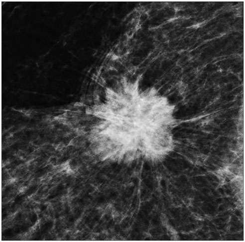
Fig. 1 Luminal A breast cancer (Ki67 = 10%, HER2 negative) presenting as mass with representative spiculated margin on mammogram. HER2 = human epithelial growth factor receptor 2
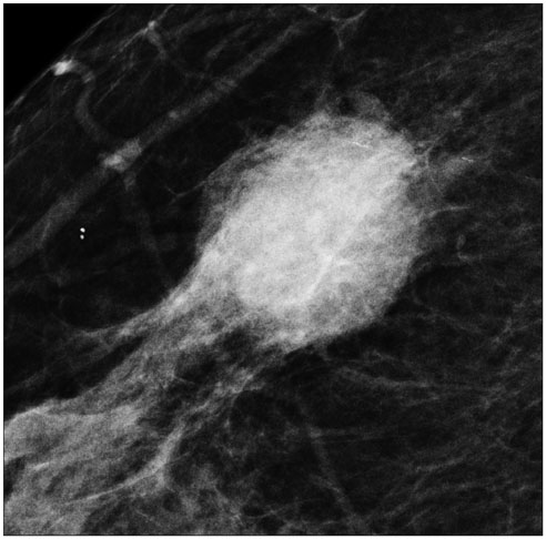
Fig. 2 Triple negative breast cancer (Ki67 = 60%, HER2 negative) presenting as mass with circumscribed margin on mammogram. HER2 = human epithelial growth factor receptor 2
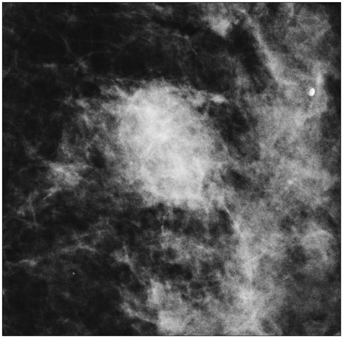
Fig. 3 HER2 breast cancer (Ki67 = 40%, HER2 positive) presenting as mass with indistinct margin on mammogram. HER2 = human epithelial growth factor receptor 2
Reference
-
1. Huber KE, Carey LA, Wazer DE. Breast cancer molecular subtypes in patients with locally advanced disease: impact on prognosis, patterns of recurrence, and response to therapy. Semin Radiat Oncol. 2009; 19:204–210.2. Lam SW, Jimenez CR, Boven E. Breast cancer classification by proteomic technologies: current state of knowledge. Cancer Treat Rev. 2014; 40:129–138.3. Guiu S, Michiels S, André F, Cortes J, Denkert C, Di Leo A, et al. Molecular subclasses of breast cancer: how do we define them? The IMPAKT 2012 Working Group Statement. Ann Oncol. 2012; 23:2997–3006.4. Jiang L, Ma T, Moran MS, Kong X, Li X, Haffty BG, et al. Mammographic features are associated with clinicopathological characteristics in invasive breast cancer. Anticancer Res. 2011; 31:2327–2334.5. Tamaki K, Ishida T, Miyashita M, Amari M, Ohuchi N, Tamaki N, et al. Correlation between mammographic findings and corresponding histopathology: potential predictors for biological characteristics of breast diseases. Cancer Sci. 2011; 102:2179–2185.6. Killelea BK, Chagpar AB, Bishop J, Horowitz NR, Christy C, Tsangaris T, et al. Is there a correlation between breast cancer molecular subtype using receptors as surrogates and mammographic appearance? Ann Surg Oncol. 2013; 20:3247–3253.7. American College of Radiology. Breast imaging reporting and data system (BI-RADS). 4th ed. Reston, VA: American College of Radiology;2003.8. Alexander MC, Yankaskas BC, Biesemier KW. Association of stellate mammographic pattern with survival in small invasive breast tumors. AJR Am J Roentgenol. 2006; 187:29–37.9. Evans AJ, Pinder SE, James JJ, Ellis IO, Cornford E. Is mammographic spiculation an independent, good prognostic factor in screening-detected invasive breast cancer? AJR Am J Roentgenol. 2006; 187:1377–1380.10. Porter GJ, Evans AJ, Cornford EJ, Burrell HC, James JJ, Lee AH, et al. Influence of mammographic parenchymal pattern in screening-detected and interval invasive breast cancers on pathologic features, mammographic features, and patient survival. AJR Am J Roentgenol. 2007; 188:676–683.11. Yamaguchi J, Ohtani H, Nakamura K, Shimokawa I, Kanematsu T. Prognostic impact of marginal adipose tissue invasion in ductal carcinoma of the breast. Am J Clin Pathol. 2008; 130:382–388.12. Moriuchi H, Yamaguchi J, Hayashi H, Ohtani H, Shimokawa I, Abiru H, et al. Cancer cell interaction with adipose tissue: correlation with the finding of spiculation at mammography. Radiology. 2016; 279:56–64.13. Tchou J, Kossenkov AV, Chang L, Satija C, Herlyn M, Showe LC, et al. Human breast cancer associated fibroblasts exhibit subtype specific gene expression profiles. BMC Med Genomics. 2012; 5:39.14. Park SY, Kim HM, Koo JS. Differential expression of cancer-associated fibroblast-related proteins according to molecular subtype and stromal histology in breast cancer. Breast Cancer Res Treat. 2015; 149:727–741.15. Baré M, Torà N, Salas D, Sentís M, Ferrer J, Ibáñez J, et al. Mammographic and clinical characteristics of different phenotypes of screen-detected and interval breast cancers in a nationwide screening program. Breast Cancer Res Treat. 2015; 154:403–415.
- Full Text Links
-
- Actions
-
Cited
- CITED
-
- Close
- Share
-
- Similar articles
-
- RE: Is There a Correlation between the Presence of a Spiculated Mass on Mammogram and Luminal A Subtype Breast Cancer?
- Detection Rate of Breast Lesion on Mammogram Shown on Breast Sonogram
- Molecular subtypes and imaging phenotypes of breast cancer
- Invasive Ductal Carcinoma vs. Invasive Lobular Carcinoma: Mammographic Findings
- Analysis of Mammographic Findings of Breast Cancer
