J Korean Ophthalmol Soc.
2019 Dec;60(12):1155-1161. 10.3341/jkos.2019.60.12.1155.
Clinical Analysis of Findings for Anterior Segment Optical Coherent Tomography in Recurrent Corneal Erosion Syndrome
- Affiliations
-
- 1Department of Ophthalmology, School of Medicine, Kyungpook National University, Daegu, Korea. okeye@knu.ac.kr
- KMID: 2466165
- DOI: http://doi.org/10.3341/jkos.2019.60.12.1155
Abstract
- PURPOSE
To evaluate the correlation between abnormal findings seen in anterior segment optical coherent tomography and the recurrence rate in patients with recurrent corneal erosion syndrome.
METHODS
Between January 2015 and August 2018, 53 eyes of 52 patients who had been diagnosed with recurrent corneal erosion syndrome were included in the study. Follow-up was performed for 12 months. To confirm the recurrence, we questioned the subjects on their symptoms and performed slit lamp examinations. At the first visit, the second week, and the first month, we performed anterior segment optical coherent tomography to identify pathologic findings for recurrent corneal erosion syndrome.
RESULTS
In 12 months, 29 eyes (54.7%) had a recurrence of corneal erosion and 24 eyes (45.3%) had no recurrence. There was no significant difference in age, sex, trauma, diabetes mellitus, or meibomian gland dysfunction between the recurrent and non-recurrent groups. Among the anterior segment optical coherent tomography findings, anterior stromal hyper-reflectivity, undetected epithelial basement membrane, intraepithelial basement membrane, intraepithelial inclusions were not significantly different between the two groups. In the first month, corneal epithelial edema was 82.8% in the recurrent group, but 33.3% in the non-recurrent group. It was significantly different (p = 0.000). Corneal epithelial thickness lowered significantly in the non-recurrent group, but not in the recurrent group in the first month. In other words, epithelial edema improved in the non-recurrent group, whereas epithelial edema did not improve in the recurrent group.
CONCLUSIONS
If corneal epithelial edema is not treated in patients with recurrent corneal erosion syndrome, high possibility of a recurrence should be considered.
Keyword
MeSH Terms
Figure
-

Figure 1 Anterior segment optical coherent tomography of theright cornea. Anterior stromal hyper-reflectivity bellow affectedepithelium (arrow), intraepithelial inclusion (arrowhead),and undetected epithelial basement membrane (asterisk).
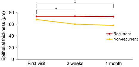
Figure 2 Change in the corneal epithelial thickness. A comparisonof the corneal epithelial thickness between the recurrentgroup and the non-recurrent group. *Wilcoxon signed rank test.
Reference
-
1. Ramamurthi S, Rahman MQ, Dutton GN, Ramaesh K. Pathogenesis, clinical features and management of recurrent corneal erosion. Eye (Lond). 2006; 20:635–644.2. Reidy JJ, Paulus MP, Gona S. Recurrent erosions of the cornea: epidemiology and treatment. Cornea. 2000; 19:767–771.3. Mandić Z, Bednar I, Šarić D. Modern approach in the treatment of recurrent corneal erosion. Acta Clin Croatica. 2007; 46:Suppl 1. 25–30.4. Suh Y, Kim MS. The longterm evaluation of recurrent corneal erosion. J Korean Ophthalmol Soc. 2002; 43:1570–1576.5. Diez-Feijóo E, Grau AE, Abusleme EI, Durán JA. Clinical presentation and causes of recurrent corneal erosion syndrome syndrome: review of 100 patients. Cornea. 2014; 33:571–575.6. Chandler PA. Recurrent erosion of the cornea. Trans Am Ophthalmol Soc. 1944; 42:355–371.7. Shin DY, Chung SH. Efficacy of anterior stromal puncture using 5% NaCl eye drops for prolonged time in recurrent corneal erosion syndrome. J Korean Ophthalmol Soc. 2017; 58:503–508.8. del Castillo JM, de la Casa JM, Sardiña RC, et al. Treatment of recurrent corneal erosion using autologous serum. Cornea. 2002; 21:781–783.9. Kwon MY, Park DJ. The clinical result of extended wear of therapeutic contact lenses and 5% NaCl for traumatic recurrent corneal erosion. J Korean Ophthalmol Soc. 2019; 60:16–24.10. Watson S, Lee H. Interventions for recurrent corneal erosion: a Cochrane systematic review. Eye (Lond). 2013; 27:1330–1331.11. Dursun D, Kim MC, Solomon A, Pflugfelder SC. Treatment of recalcitrant recurrent corneal erosion with inhibitors of matrix metalloproteinase-9, doxycycline and corticosteroids. Am J Ophthalmol. 2001; 132:8–13.12. Das S, Seitz B. Recurrent corneal erosion syndrome. Surv Ophthalmol. 2008; 53:3–15.13. McLean EN, MacRae SM, Rich LF. Recurrent erosion. Treatment by anterior stromal puncture. Ophthalmology. 1986; 93:784–788.14. Soong HK, Farjo Q, Meyer RF, Sugar A. Diamond burr superficial keratectomy for recurrent corneal erosions. Br J Ophthalmol. 2002; 86:296–298.15. Ohman L, Fagerholm P. The influence of excimer laser ablation on recurrent corneal erosion: a prospective randomized study. Cornea. 1998; 17:349–352.16. Bea KH, Ahn M, Cho NC, You IC. Clinical presentation and treatment outcomes of recurrent corneal erosion. J Korean Ophthalmol Soc. 2016; 57:555–561.17. Korea External Eye Disease Society. Cornea. 3rd ed. Seoul: :Ilchokak;2013. p. 297–302.18. Diez-Feijóo E, Durán JA. Optical coherence tomography findings in recurrent corneal erosion syndrome. Cornea. 2015; 34:290–295.19. Rocha KM, Perez-Straziota CE, Stulting RD, Randleman JB. SD-OCT analysis of regional epithelial thickness profiles in keratoconus, postoperative corneal ectasia, and normal eyes. J Refract Surg. 2013; 29:173–179.20. Reinstein DZ, Archer TJ, Gobbe M, et al. Epithelial thickness in the normal cornea: three-dimensional display with Artemis very high-frequency digital ultrasound. J Refract Surg. 2008; 24:571–581.21. Feng Y, Simpson TL. Corneal, limbal, and conjunctival epithelial thickness from optical coherence tomography. Optom Vis Sci. 2008; 85:E880–E883.22. Gipson IK, Spurr-Michaud SJ, Tisdale AS. Anchoring fibrils form a complex network in human and rabbit cornea. Invest Ophthalmol Vis Sci. 1987; 28:212–220.23. Wood TO, Judge D, Payant J, Frase S. Electron microscopy of anterior stromal micropuncture. Invest Ophthalmol Vis Sci. 1989; 30:Suppl. 1.24. Garrana RM, Zieske JD, Assouline M, Gipson IK. Matrix metalloproteinases in epithelia from human recurrent corneal erosion. Invest Ophthalmol Vis Sci. 1999; 40:1266–1270.25. Hykin PG, Foss AE, Pavesio C, Dart JK. The natural history and management of recurrent corneal erosion: a prospective randomized trial. Eye (Lond). 1994; 8 Pt 1:35–40.26. Choi M, Jung JW, Seo KY, et al. Comparison of Nd:YAG laser versus conservative management in the treatment of recurrent corneal erosion. J Korean Ophthalmol Soc. 2015; 56:687–693.27. Brown N, Bron A. Recurrent erosion of the cornea. Br J Ophthalmol. 1976; 60:84–96.
- Full Text Links
-
- Actions
-
Cited
- CITED
-
- Close
- Share
-
- Similar articles
-
- Comparison of Anterior Segment Parameters Obtained by Anterior Segment Optical Coherence Tomography and Dual Rotating Scheimpflug Camera
- Utility of the Anterior Segment Optical Coherence Tomography for Measurements of Central Corneal Thickness
- Refractory Recurrent Corneal Erosion after Descemet’s Stripping Automated Endothelial Keratoplasty
- Therapeutic Effect of Recurrent Corneal Erosion by Nd: YAG Anterior Stromal Puncture
- Changes in Corneal Thickness in Congenital Glaucoma, Using Anterior Segment Optical Coherence Tomography
