A Novel Immunomodulatory Mechanism Dependent on Acetylcholine Secreted by Human Bone Marrow-derived Mesenchymal Stem Cells
- Affiliations
-
- 1Department of Integrated Biomedical Sciences, Inha University School of Medicine, Incheon, Korea. sunuksong@inha.ac.kr
- 2SCM Lifescience Co., Ltd., Incheon, Korea.
- 3SunCreate Co., Ltd., Yangju, Korea.
- 4Department of Radiooncology, Inha University School of Medicine, Incheon, Korea.
- KMID: 2465901
- DOI: http://doi.org/10.15283/ijsc18098
Abstract
- BACKGROUND AND OBJECTIVES
Mesenchymal stem cells (MSCs) are used to treat autoimmune or inflammatory diseases. Our aim was to determine the immunomodulatory mechanisms elicited by MSCs during inflammation.
METHODS AND RESULTS
We cocultured MSCs with peripheral blood mononuclear cells for a mixed lymphocyte reaction or stimulated them by phytohemagglutinin. Morphological changes of MSCs and secretion of acetylcholine (ACh) from MSCs were measured. The effects of an ACh antagonist and ACh agonist on lymphocyte proliferation and proinflammatory-cytokine production were determined. The inflammatory milieu created by immune-cell activation caused MSCs to adopt a neuronlike phenotype and induced them to release ACh. Additionally, nicotinic acetylcholine receptors (nAChRs) were upregulated in activated peripheral blood mononuclear cells. We observed that ACh bound to nAChR on activated immune cells and led to the inhibition of lymphocyte proliferation and of proinflammatory-cytokine production. MSC-mediated immunosuppression through ACh activity was reversed by an ACh antagonist called α-bungarotoxin, and lymphocyte proliferation was inhibited by an ACh agonist, ACh chloride.
CONCLUSIONS
Our findings point to a novel immunomodulatory mechanism in which ACh secreted by MSCs under inflammatory conditions might modulate immune cells. This study may provide a novel method for the treatment of autoimmune diseases by means of MSCs.
MeSH Terms
Figure
-

Fig. 1 Inflammatory conditions induce neuronlike morphological features in MSCs. (a) When cocultured in an MLR with PBMCs obtained from two individuals (P and Po), hBM MSCs suppressed the lymphocyte proliferation. (b) When cocultured with human PBMCs activated by PHA stimulation (PPHA), MSCs inhibited the lymphocyte proliferation. CPM: counts per million. (c) Morphological changes in MSCs were observed in cocultures with activated PBMCs. (d, e) Neuronlike morphological changes in MSCs after coculture with activated PBMCs for 48 h. (f, g) Floating neurosphere-like cell clusters (red arrows) were observed under both inflammatory conditions. Error bars are indicative of standard deviations.
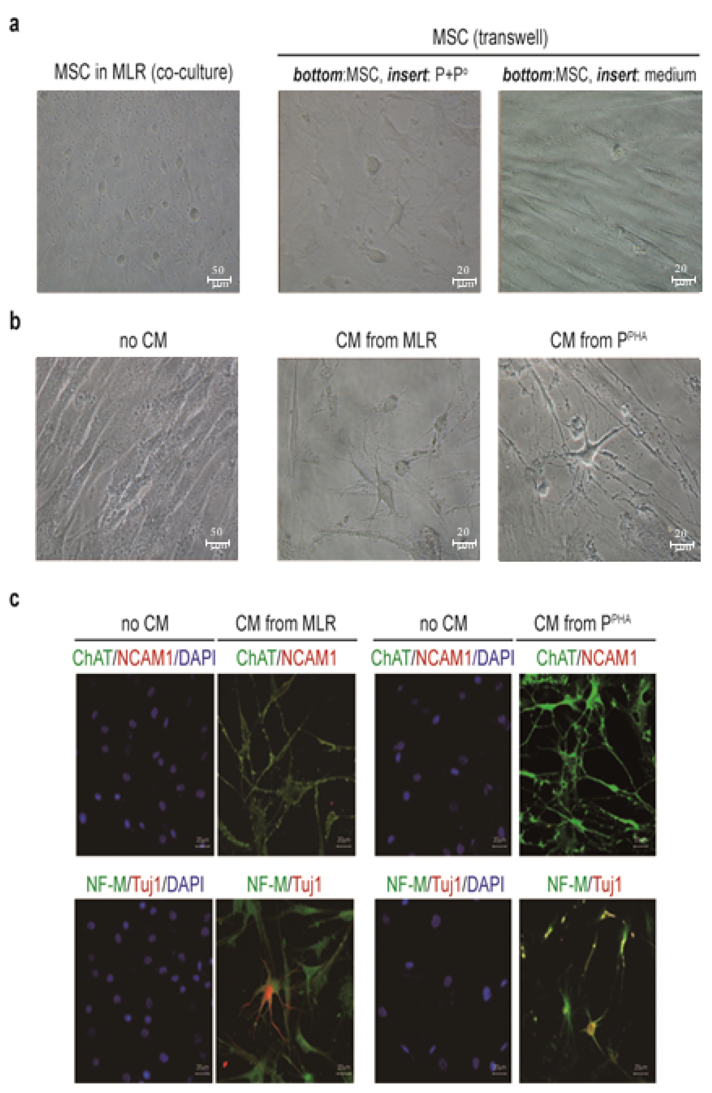
Fig. 2 Inflammation induces the expression of NRs and neuron- or NPC-related markers but not glial markers on the MSC surface. (a) To determine whether inflammation-induced neuronlike phenotypic changes in MSCs were mediated by soluble factors, we performed assays using Transwell plates with inserts (0.4 μm pore size). MSCs (105) were seeded in the bottom well, and MLR-activated PBMCs (106) were seeded in the insert-containing well. After incubation for 3 days, MSCs at the bottom went through neuronlike morphological changes. These changes were not observed in the absence of activated PBMCs in the insert. (b) When MSCs were incubated for 3 days with the CM from activated PBMCs, they acquired neuronlike morphology. (c) MSCs incubated with the CM from activated PBMCs expressed ChAT, NCAM1, NF-M, and TUJ1.

Fig. 3 Inflammation induces the expression of NRs and neuron- or NPC-related markers but not glial markers on the MSC surface. (a) RT-PCR was carried out to assess the expression of nestin, Tuj1, MAP2, NF-M, and GFAP in adherent MSCs cocultured with activated PBMCs (24 h). GAPDH served as a loading control. (b) Flow-cytometric analysis of TUJ1, nestin, and GFAP in adherent MSCs cocultured with activated PBMCs (24 h). (c) Immunofluorescence staining of nestin, TUJ1, NCAM1, GFAP, and O4 in MSCs cocultured in the MLR for 48 h. (d) Immunofluorescence staining for nestin, TUJ1, NCAM1, GFAP, and O4 in MSCs cocultured with PHA-activated PBMCs for 48 h. DAPI was used to stain nuclei. (e) RT-PCR analysis of TrkA, TrkB, TrkC, and p75NTR expression in adherent MSCs cocultured with activated PBMCs (24 h). Tuj1 served as a control for an MSC response to inflammation, and GAPDH was employed as a loading control. (f, g) qPCR was conducted to assess the expression of the aforementioned NRs as in (e). (h) Protein expression of TrkA and p75NTR, but not TrkC, was validated by western blotting in the whole MSC extracts (20 μg) after 48 h of coculture. (i, j) TrkA induction in MSCs in response to experimental inflammation was confirmed by immunofluorescence staining. Error bars are indicative of standard deviations.

Fig. 4 Neurotrophins are induced in PBMCs activated by MLR or PHA. (a) RT-PCR analysis of NGF and BDNF expression in PBMCs after activation by MLR for 24 h. (b) qPCR analysis of NGF and BDNF expression in the samples as described in panel (a). (c) RT-PCR analysis of NGF and BDNF expression in PBMCs after stimulation with PHA for 24 h. (d) qPCR analysis of NGF and BDNF expression in the samples as described in panel (c). (e~h) ELISA quantification of soluble NGF and BDNF in the CM obtained from activated-PBMC cultures (48 h). Three independent experiments were conducted.
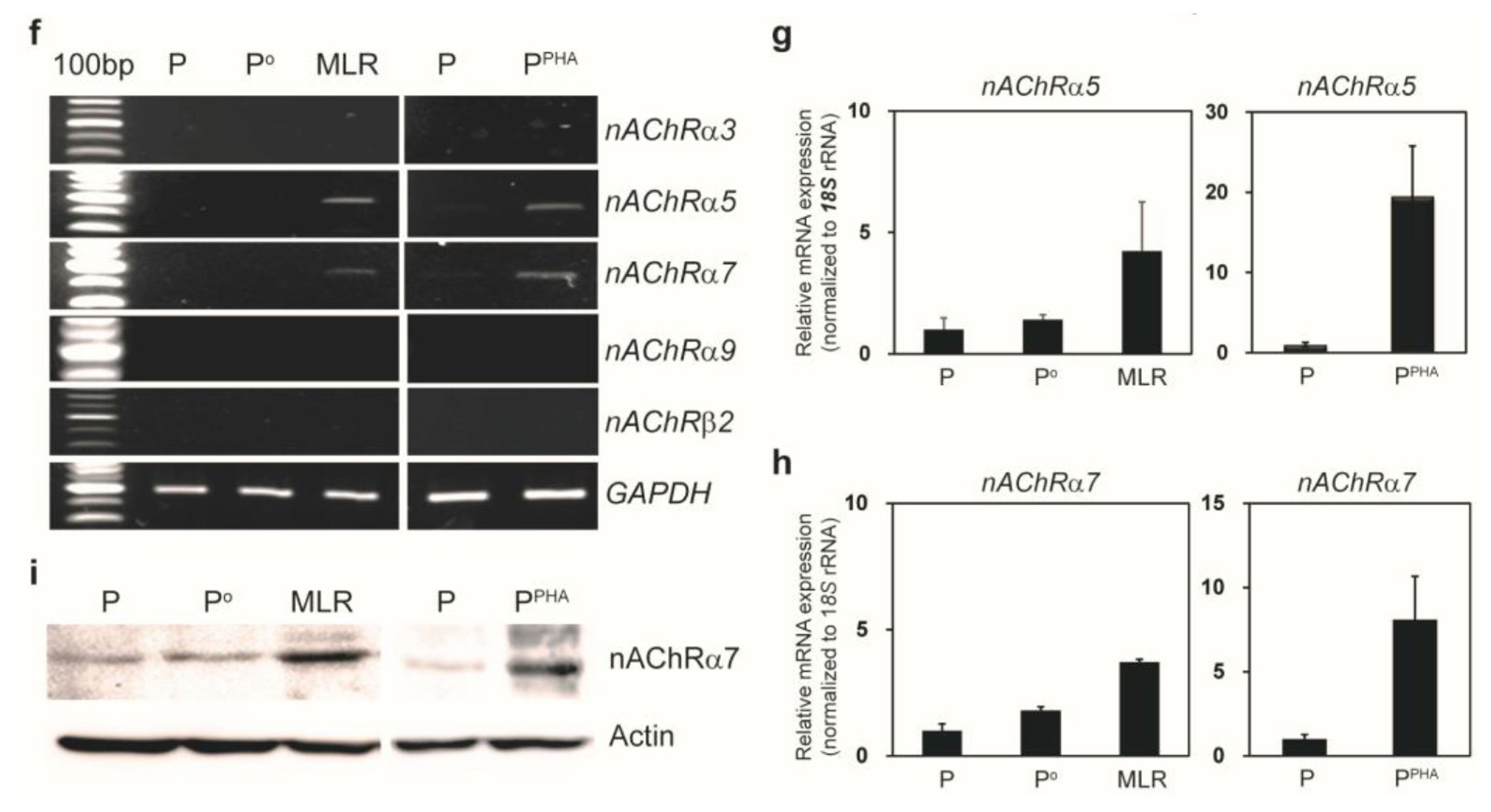
Fig. 5 Inflammatory conditions induce a cholinergic-neuron–like phenotype in MSCs and nAChRs in activated PBMCs. (a) Immunofluorescence staining of ChAT, GABA, and TH in MSCs cocultured with activated PBMCs (48 h). (b) qPCR was carried out to quantify ChAT expression in MSCs after inflammatory stimulation for 24 h. (c) Western blotting confirmed ChAT expression in the whole MSC extract (20 μg) after coculture for 48 h. (d) ACh and choline concentration was measured in the CM obtained from PBMCs alone (P or Po), PHA-activated PBMCs (PPHA), or MLR (P and Po) culture without or with MSCs (n=3). (e) ACh and choline concentration was measured in the CM obtained from PBMCs alone (P or Po) or MLR (P and Po) culture without or with MSCs for 48 h. “MLR sup.” is the supernatant from the MLR without MSCs. “(MLR+MSC) sup.” is the supernatant from MLR with MSCs (n=3). (f) RT-PCR analysis of several nAChR subunits in activated PBMCs. (g) qPCR was carried out to assess nAChR α5 expression in activated PBMCs after incubation for 24 h. (h) qPCR was performed to measure nAChR α7 expression in activated PBMCs after incubation for 24 h. (i) An increase in the protein expression of the nAChR α7 subunit in activated PBMCs was confirmed by western blotting of whole MSC extracts prepared after MLR or PHA stimulation for 48 h.
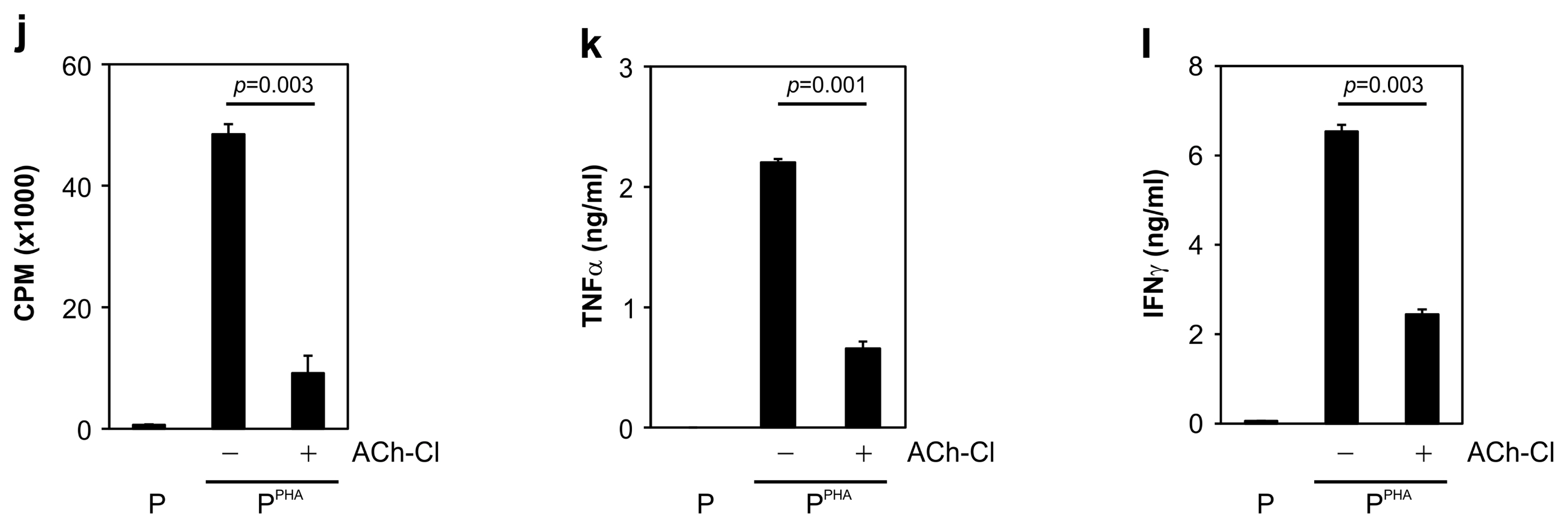
Fig. 6 MSC-mediated immunosuppression via ACh is reversed by α-BTX (ACh antagonist), and lymphocyte proliferation is inhibited by ACh-Cl (ACh agonist). (a) MSC-suppressed lymphocyte proliferation was significantly restored by α-BTX addition to the MLR medium. (b) MSC-suppressed TNF-α production during MLR was restored by α-BTX treatment. (c) Suppressed secretion of IFN-γ during MLR was significantly restored by α-BTX treatment. (d) MSC-mediated suppression of PHA-stimulated lymphocyte proliferation was significantly attenuated by α-BTX treatment. (e) MSC-suppressed TNF-α production in PHA-activated PBMCs increased in the presence of α-BTX. (f) The reduced IFN-γ secretion by PHA-activated PBMCs was restored by α-BTX. (g, j) ACh-Cl addition to the medium significantly attenuated the increase in lymphocyte proliferation caused by (g) MLR or (j) PHA treatment. (h, k) Production of TNF-α by activated PBMCs elicited by (h) MLR or (k) PHA stimulation was attenuated by ACh-Cl treatment. (i, l) ACh-Cl treatment significantly suppressed the IFN-γ secretion from PBMCs activated by (i) MLR or (l) PHA stimulation. All data are the average of 3~5 independent experiments and were statistically evaluated by paired Student’s t test. p values <0.05 were assumed to indicate statistically significant variations.
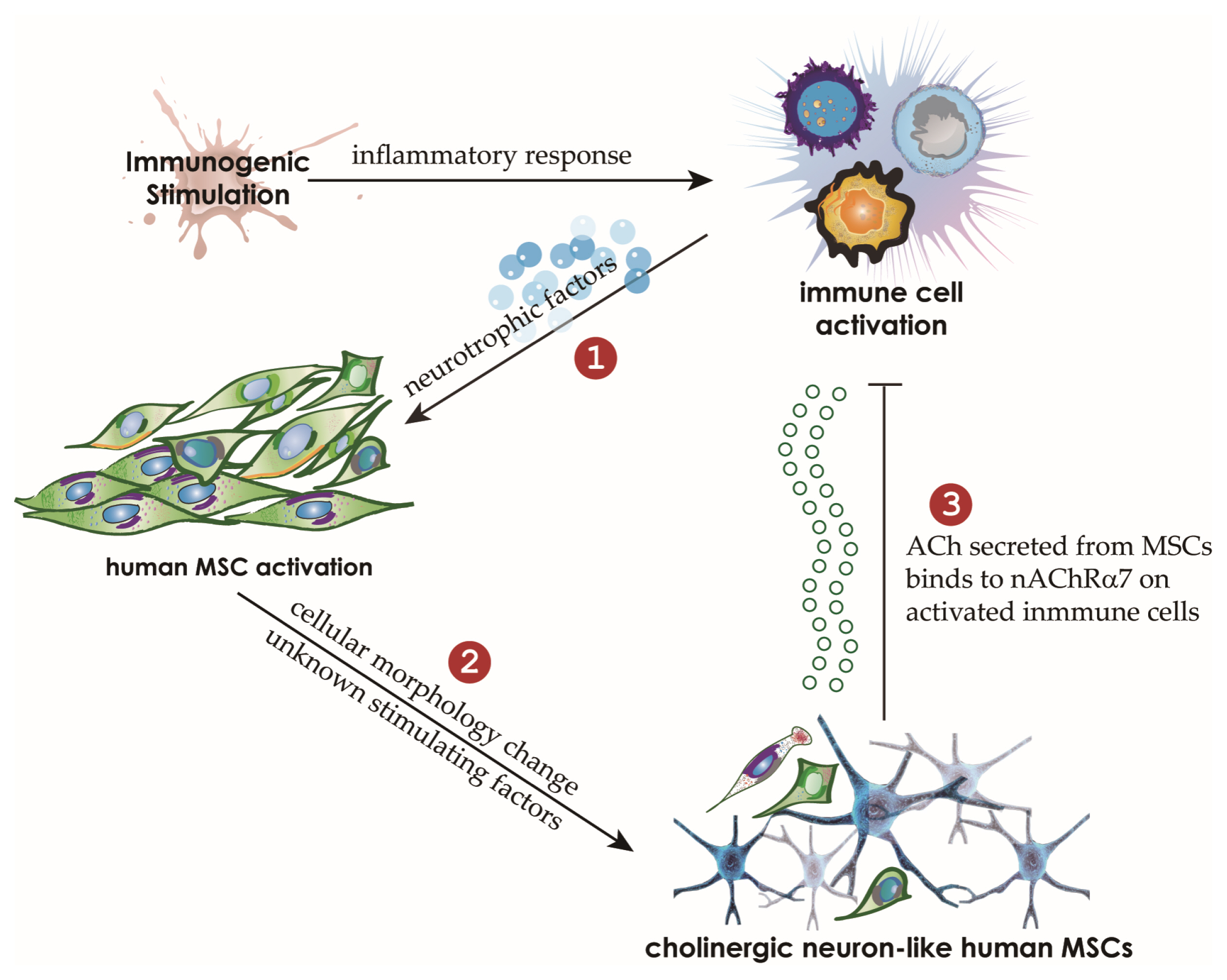
Fig. 7 The proposed model of an immunomodulatory mechanism utilized by human MSCs in an inflammatory milieu. The inflammatory conditions drive human MSCs to adopt a neuronlike phenotype. It is probable that the expression of neurotrophins such as NGF and BDNF in human AICs and the presence of NRs on MSCs are associated with these changes in MSCs. Furthermore, the inflammatory milieu probably induces the expression of nAChR including nAChR α7, which participates in the negative regulation of activated lymphocytes. Neuronlike MSCs stimulated by neurotrophins and unknown factors secrete ACh, which binds to AICs via nAChR α7, thereby inhibiting the proliferation and function of AICs.
Cited by 1 articles
-
Perivascular Stem Cells Suppress Inflammasome Activation during Inflammatory Responses in Macrophages
Jeeyoung Kim, Woo Jin Kim, Kwon-Soo Ha, Eun-Taek Han, Won Sun Park, Se-Ran Yang, Seok-Ho Hong
Int J Stem Cells. 2019;12(3):419-429. doi: 10.15283/ijsc19115.
Reference
-
References
1. Dzierzak E, Speck NA. Of lineage and legacy: the development of mammalian hematopoietic stem cells. Nat Immunol. 2008; 9:129–136. DOI: 10.1038/ni1560. PMID: 18204427. PMCID: PMC2696344.
Article2. Pittenger MF, Mackay AM, Beck SC, Jaiswal RK, Douglas R, Mosca JD, Moorman MA, Simonetti DW, Craig S, Marshak DR. Multilineage potential of adult human mesenchymal stem cells. Science. 1999; 284:143–147. DOI: 10.1126/science.284.5411.143. PMID: 10102814.
Article3. Charbord P. Bone marrow mesenchymal stem cells: historical overview and concepts. Hum Gene Ther. 2010; 21:1045–1056. DOI: 10.1089/hum.2010.115. PMID: 20565251. PMCID: PMC4823383.
Article4. Scuteri A, Miloso M, Foudah D, Orciani M, Cavaletti G, Tredici G. Mesenchymal stem cells neuronal differentiation ability: a real perspective for nervous system repair? Curr Stem Cell Res Ther. 2011; 6:82–92. DOI: 10.2174/157488811795495486. PMID: 21190538.
Article5. Fischer UM, Harting MT, Jimenez F, Monzon-Posadas WO, Xue H, Savitz SI, Laine GA, Cox CS Jr. Pulmonary passage is a major obstacle for intravenous stem cell delivery: the pulmonary first-pass effect. Stem Cells Dev. 2009; 18:683–692. DOI: 10.1089/scd.2008.0253. PMID: 19099374. PMCID: PMC3190292.
Article6. De Miguel MP, Fuentes-Julián S, Blázquez-Martínez A, Pascual CY, Aller MA, Arias J, Arnalich-Montiel F. Immunosuppressive properties of mesenchymal stem cells: advances and applications. Curr Mol Med. 2012; 12:574–591. DOI: 10.2174/156652412800619950. PMID: 22515979.
Article7. Kode JA, Mukherjee S, Joglekar MV, Hardikar AA. Mesenchymal stem cells: immunobiology and role in immunomodulation and tissue regeneration. Cytotherapy. 2009; 11:377–391. DOI: 10.1080/14653240903080367. PMID: 19568970.
Article8. Le Blanc K, Frassoni F, Ball L, Locatelli F, Roelofs H, Lewis I, Lanino E, Sundberg B, Bernardo ME, Remberger M, Dini G, Egeler RM, Bacigalupo A, Fibbe W, Ringdén O. Mesenchymal stem cells for treatment of steroid-resistant, severe, acute graft-versus-host disease: a phase II study. Lancet. 2008; 371:1579–1586. DOI: 10.1016/S0140-6736(08)60690-X. PMID: 18468541.
Article9. Parekkadan B, Upadhyay R, Dunham J, Iwamoto Y, Mizoguchi E, Mizoguchi A, Weissleder R, Yarmush ML. Bone marrow stromal cell transplants prevent experimental enterocolitis and require host CD11b+ splenocytes. Gastroenterology. 2011; 140:966–975. DOI: 10.1053/j.gastro.2010.10.013. PMCID: PMC3033974.
Article10. Jung KH, Song SU, Yi T, Jeon MS, Hong SW, Zheng HM, Lee HS, Choi MJ, Lee DH, Hong SS. Human bone marrow-derived clonal mesenchymal stem cells inhibit inflammation and reduce acute pancreatitis in rats. Gastroenterology. 2011; 140:998–1008. DOI: 10.1053/j.gastro.2010.11.047. PMID: 21130088.
Article11. Na K, Yoo HS, Zhang YX, Choi MS, Lee K, Yi TG, Song SU, Jeon MS. Bone marrow-derived clonal mesenchymal stem cells inhibit ovalbumin-induced atopic dermatitis. Cell Death Dis. 2014; 5:e1345. DOI: 10.1038/cddis.2014.299. PMID: 25032868. PMCID: PMC4123091.
Article12. Stagg J, Galipeau J. Mechanisms of immune modulation by mesenchymal stromal cells and clinical translation. Curr Mol Med. 2013; 13:856–867. DOI: 10.2174/1566524011313050016. PMID: 23642066.
Article13. Soleymaninejadian E, Pramanik K, Samadian E. Immunomodulatory properties of mesenchymal stem cells: cytokines and factors. Am J Reprod Immunol. 2012; 67:1–8. DOI: 10.1111/j.1600-0897.2011.01069.x. PMID: 21951555.
Article14. Sternberg EM. Neural regulation of innate immunity: a coordinated nonspecific host response to pathogens. Nat Rev Immunol. 2006; 6:318–328. DOI: 10.1038/nri1810. PMID: 16557263. PMCID: PMC1783839.
Article15. Lukewich MK, Rogers RC, Lomax AE. Divergent neuroendocrine responses to localized and systemic inflammation. Semin Immunol. 2014; 26:402–408. DOI: 10.1016/j.smim.2014.01.004. PMID: 24486057. PMCID: PMC4128895.
Article16. Padro CJ, Sanders VM. Neuroendocrine regulation of inflammation. Semin Immunol. 2014; 26:357–368. DOI: 10.1016/j.smim.2014.01.003. PMID: 24486056. PMCID: PMC4116469.
Article17. Andersson U, Tracey KJ. Reflex principles of immunological homeostasis. Annu Rev Immunol. 2012; 30:313–335. DOI: 10.1146/annurev-immunol-020711-075015. PMID: 22224768. PMCID: PMC4533843.
Article18. Kawashima K, Fujii T. The lymphocytic cholinergic system and its biological function. Life Sci. 2003; 72:2101–2109. DOI: 10.1016/S0024-3205(03)00068-7. PMID: 12628464.
Article19. Sato KZ, Fujii T, Watanabe Y, Yamada S, Ando T, Kazuko F, Kawashima K. Diversity of mRNA expression for muscarinic acetylcholine receptor subtypes and neuronal nicotinic acetylcholine receptor subunits in human mononuclear leukocytes and leukemic cell lines. Neurosci Lett. 1999; 266:17–20. DOI: 10.1016/S0304-3940(99)00259-1. PMID: 10336173.
Article20. Pavlov VA, Wang H, Czura CJ, Friedman SG, Tracey KJ. The cholinergic anti-inflammatory pathway: a missing link in neuroimmunomodulation. Mol Med. 2003; 9:125–134. DOI: 10.1007/BF03402177. PMID: 14571320. PMCID: PMC1430829.
Article21. Wang H, Yu M, Ochani M, Amella CA, Tanovic M, Susarla S, Li JH, Wang H, Yang H, Ulloa L, Al-Abed Y, Czura CJ, Tracey KJ. Nicotinic acetylcholine receptor alpha7 subunit is an essential regulator of inflammation. Nature. 2003; 421:384–388. DOI: 10.1038/nature01339. PMID: 12508119.
Article22. Fujii T, Takada-Takatori Y, Kawashima K. Basic and clinical aspects of non-neuronal acetylcholine: expression of an independent, non-neuronal cholinergic system in lymphocytes and its clinical significance in immunotherapy. J Pharmacol Sci. 2008; 106:186–192. DOI: 10.1254/jphs.FM0070109. PMID: 18285654.
Article23. Fujii YX, Fujigaya H, Moriwaki Y, Misawa H, Kasahara T, Grando SA, Kawashima K. Enhanced serum antigen-specific IgG1 and proinflammatory cytokine production in nicotinic acetylcholine receptor alpha7 subunit gene knockout mice. J Neuroimmunol. 2007; 189:69–74. DOI: 10.1016/j.jneuroim.2007.07.003. PMID: 17675251.
Article24. Hoogduijn MJ, Cheng A, Genever PG. Functional nicotinic and muscarinic receptors on mesenchymal stem cells. Stem Cells Dev. 2009; 18:103–112. DOI: 10.1089/scd.2008.0032. PMID: 18393628.
Article25. Hendrickson ML, Rao AJ, Demerdash ON, Kalil RE. Expression of nestin by neural cells in the adult rat and human brain. PLoS One. 2011; 6:e18535. DOI: 10.1371/journal.pone.0018535. PMID: 21490921. PMCID: PMC3072400.
Article26. Chao MV. Neurotrophins and their receptors: a convergence point for many signalling pathways. Nat Rev Neurosci. 2003; 4:299–309. DOI: 10.1038/nrn1078. PMID: 12671646.
Article27. Lambiase A, Bracci-Laudiero L, Bonini S, Bonini S, Starace G, D’Elios MM, De Carli M, Aloe L. Human CD4+ T cell clones produce and release nerve growth factor and express high-affinity nerve growth factor receptors. J Allergy Clin Immunol. 1997; 100:408–414. DOI: 10.1016/S0091-6749(97)70256-2. PMID: 9314355.
Article28. Kawashima K, Fujii T. Extraneuronal cholinergic system in lymphocytes. Pharmacol Ther. 2000; 86:29–48. DOI: 10.1016/S0163-7258(99)00071-6. PMID: 10760545.
Article29. Grando SA, Kawashima K, Wessler I. Introduction: the non-neuronal cholinergic system in humans. Life Sci. 2003; 72:2009–2012. DOI: 10.1016/S0024-3205(03)00063-8. PMID: 12628450.
Article30. Borovikova LV, Ivanova S, Zhang M, Yang H, Botchkina GI, Watkins LR, Wang H, Abumrad N, Eaton JW, Tracey KJ. Vagus nerve stimulation attenuates the systemic inflammatory response to endotoxin. Nature. 2000; 405:458–462. DOI: 10.1038/35013070. PMID: 10839541.
Article31. Kawashima K, Yoshikawa K, Fujii YX, Moriwaki Y, Misawa H. Expression and function of genes encoding cholinergic components in murine immune cells. Life Sci. 2007; 80:2314–2319. DOI: 10.1016/j.lfs.2007.02.036. PMID: 17383684.
Article32. Saeed RW, Varma S, Peng-Nemeroff T, Sherry B, Balakhaneh D, Huston J, Tracey KJ, Al-Abed Y, Metz CN. Cholinergic stimulation blocks endothelial cell activation and leukocyte recruitment during inflammation. J Exp Med. 2005; 201:1113–1123. DOI: 10.1084/jem.20040463. PMID: 15809354. PMCID: PMC2213139.
Article33. Nizri E, Hamra-Amitay Y, Sicsic C, Lavon I, Brenner T. Anti-inflammatory properties of cholinergic up-regulation: a new role for acetylcholinesterase inhibitors. Neuropharmacology. 2006; 50:540–547. DOI: 10.1016/j.neuropharm.2005.10.013. PMID: 16336980.
Article34. Shen JX, Yakel JL. Nicotinic acetylcholine receptor-mediated calcium signaling in the nervous system. Acta Pharmacol Sin. 2009; 30:673–680. DOI: 10.1038/aps.2009.64. PMID: 19448647. PMCID: PMC4002362.
Article35. Fauchais AL, Boumediene A, Lalloue F, Gondran G, Loustaud-Ratti V, Vidal E, Jauberteau MO. Brain-derived neurotrophic factor and nerve growth factor correlate with T-cell activation in primary Sjogren’s syndrome. Scand J Rheumatol. 2009; 38:50–57. DOI: 10.1080/03009740802378832. PMID: 18830907.
Article36. Ehrhard PB, Erb P, Graumann U, Otten U. Expression of nerve growth factor and nerve growth factor receptor tyrosine kinase Trk in activated CD4-positive T-cell clones. Proc Natl Acad Sci U S A. 1993; 90:10984–10988. DOI: 10.1073/pnas.90.23.10984. PMID: 7902578. PMCID: PMC47906.
Article
- Full Text Links
-
- Actions
-
Cited
- CITED
-
- Close
- Share
-
- Similar articles
-
- Immunomodulatory Effects of Placenta-derived Mesenchymal Stem Cells on T Cells by Regulation of FoxP3 Expression
- Clinical Safety and Efficacy of Autologous Bone Marrow-Derived Mesenchymal Stem Cell Transplantation in Sensorineural Hearing Loss Patients
- Concise Review: Differentiation of Human Adult Stem Cells Into Hepatocyte-like Cells In vitro
- Immunomodulatory Effect of Epidermal Growth Factor Secreted by Human Umbilical Cord Blood-Derived Mesenchymal Stem Cells on Atopic Dermatitis
- Bone marrow-derived stem cells contribute to regeneration of the endometrium
