Characterization and Differentiation of Circulating Blood Mesenchymal Stem Cells and the Role of Phosphatidylinositol 3-Kinase in Modulating the Adhesion
- Affiliations
-
- 1Dental Research Institute, Seoul National University, Brain Korea 21, Seoul, Korea.
- 2Dental Research Institute and Prosthodontics, Seoul National University Dental Hospital, School of Dentistry, Seoul National University, Seoul, Korea. ksy0617@snu.ac.kr
- 3Department of Dental Regenerative Biotechnology, School of Dentistry, Seoul National University, Seoul, Korea. jcho@snu.ac.kr
- KMID: 2465897
- DOI: http://doi.org/10.15283/ijsc18136
Abstract
- Bone marrow mesenchymal stem cells (BM MSCs) can differentiate into multi-lineage tissues. However, obtaining BM MSCs by aspiration is difficult and can be painful; therefore peripheral blood (PB) MSCs might provide an easier alternative for clinical applications. Here, we show that circulating PB MSCs proliferate as efficiently as BM MSCs in the presence of extracellular matrix (ECM) and that differentiation potential into osteoblast in vitro and in vivo. Both BM MSCs and PB MSCs developed into new bone when subcutaneously transplanted into immune-compromised mice using hydroxyapatite/tricalcium phosphate as a carrier. Furthermore, LY294002 and Wortmannin blocked mesenchymal stem cell attachment in a dose-dependent manner, suggesting a role of phosphatidylinositol 3-kinase in MSC attachment. Our data showed that the growth of PB MSCs could be regulated by interaction with the ECM and that these cells could differentiate into osteoblasts, suggesting their potential for clinical applications.
Keyword
MeSH Terms
Figure
-
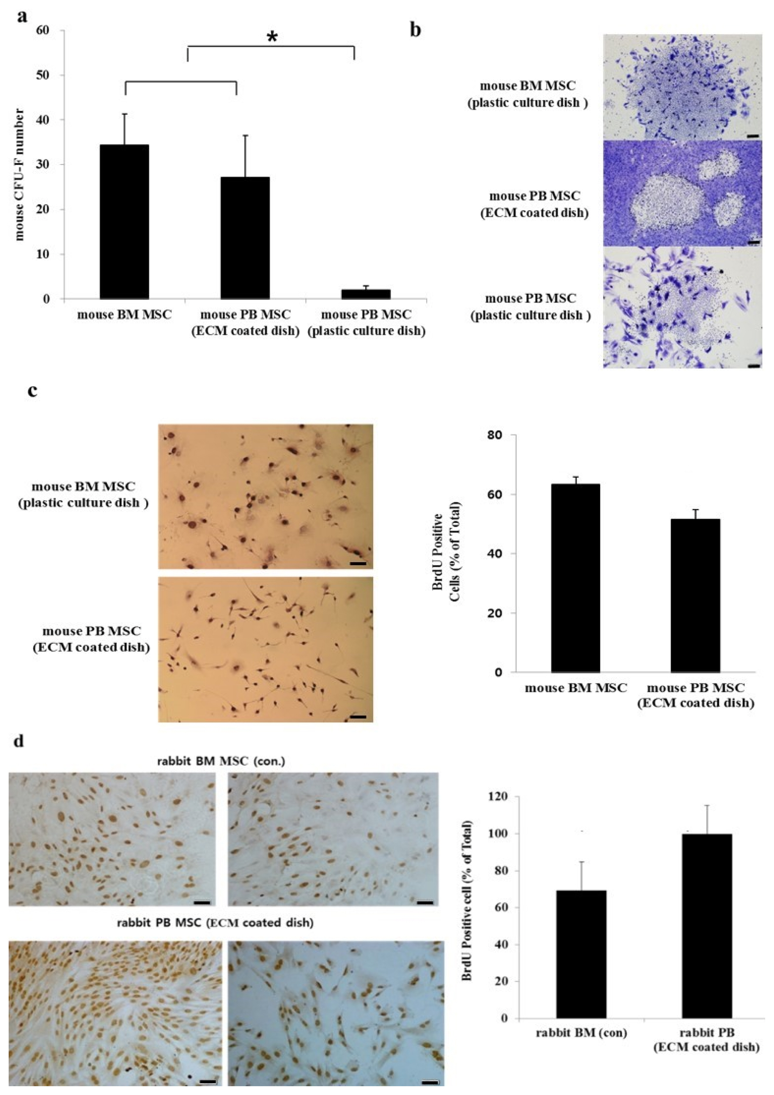
Fig. 1 CFU-F and proliferation of BM MSCs and PB MSCs. (a) Mouse BM MSCs seeded on the normal culture dish showed substantial proliferation whereas mouse PB MSCs seeded on the same kind of dish produced significantly fewer CFU-Fs (*p<0.05). There was a significant increase in the number of mouse PB MSCs plated on ECM-coated culture dish compared with mouse PB MSCs plated on a normal dish (*p<0.05). (b) Analysis of colony morphology of three dishes at ×40 magnification revealed that BM MSCs adhere to each other in contrast to the dispersed nature of mouse PB MSCs in culture. (c) Self-renewal capacity of mouse BM MSCs and mouse PB MSCs and morphology of these cells (×200 magnification). The proliferation of mouse BM MSCs and mouse PB MSCs measured by BrdU incorporation were similar (*p>0.05). Data were obtained from the mean±SE of nine fields. (d) Self-renewal of rabbit BM MSCs and rabbit PB MSCs. The proliferation of rBM MSCs and rPB MSCs measured by BrdU incorporation. Analysis of variance (**p<0.01). Scale bars=50 μM.
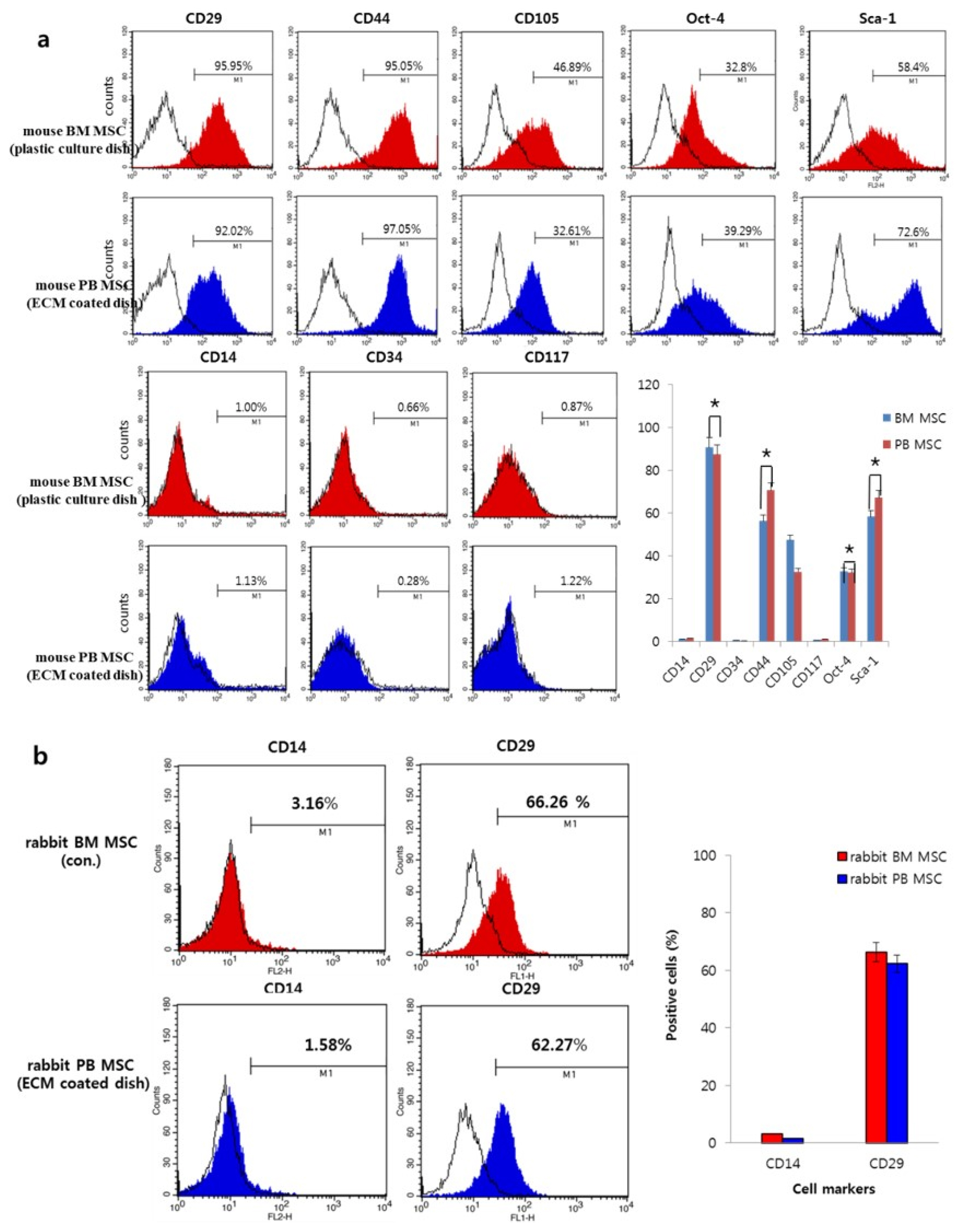
Fig. 2 FACS analysis. (a) Flow cytometric analysis of the expression of mouse markers related to stem cells such as MSCs and hematopoietic stem cells. (b) Flow cytometric analysis of the expression of rabbit cell markers related to stem cells such as MSCs and hematopoietic stem cells. Data show mean±SE of three independent experiments (*p>0.05).
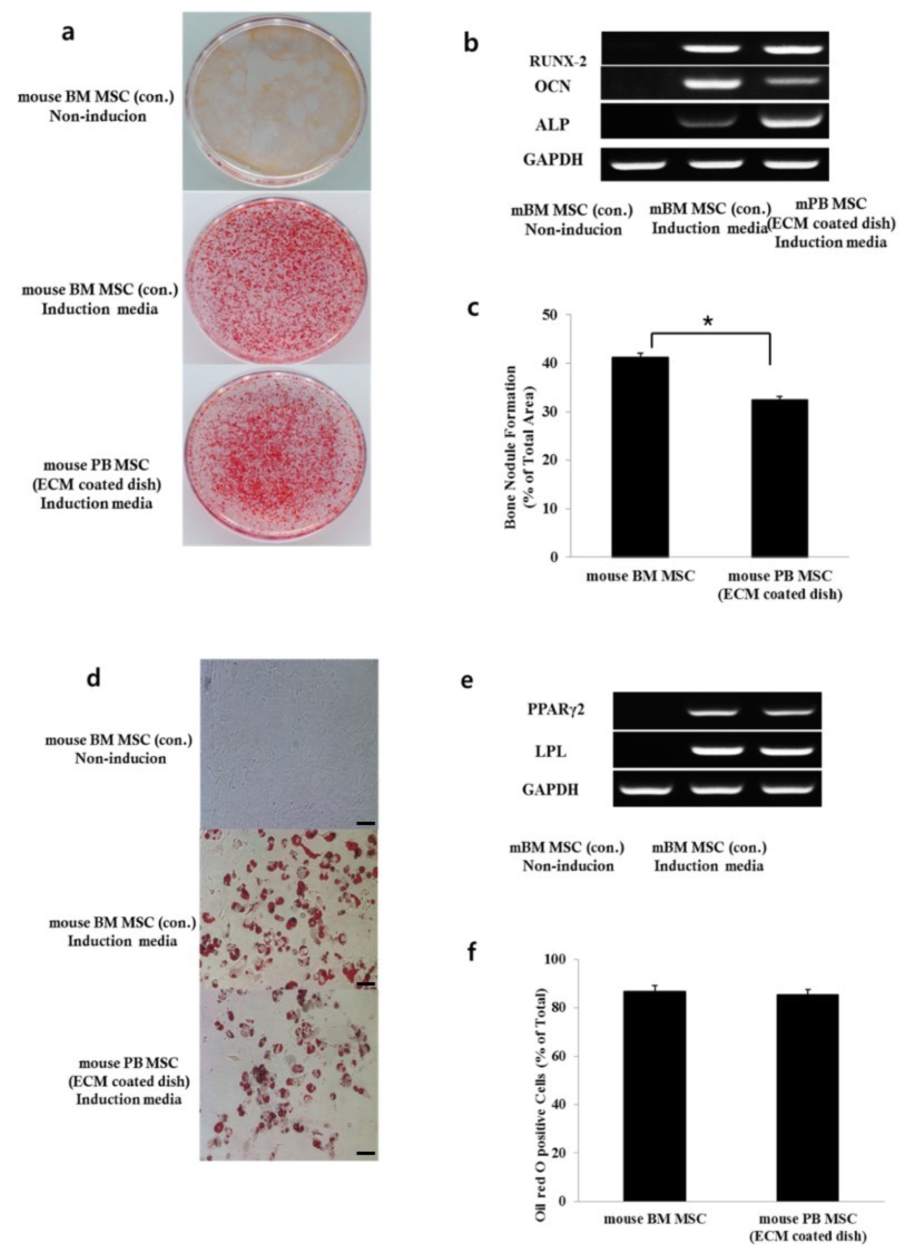
Fig. 3 Osteogenic and adipogenic differentiation of mouse BM MSCs and mouse PB MSCs in vitro. (a) Calcium accumulation revealed by alizarin red S staining at ×200 magnification. (b) Expression of RUNX-2, OCN, and ALP, which induce osteoblast differentiation, in both experimental groups. (c) Percentage of mineralized area/total area of the dish. Data show mean±SE of three dishes (*p<0.05). (d) Lipid droplets revealed by Oil red O staining at ×200 magnification indicating that the MSCs are capable of forming Oil red O-positive cells. (e) RT-PCR showing positive gene expression profiles related to adipogenic differentiation in induced cultures compared with uninduced (un) cultures. (f) A number of cells staining positive/total number of cells. Data show mean±SE of five fields (*p>0.05).
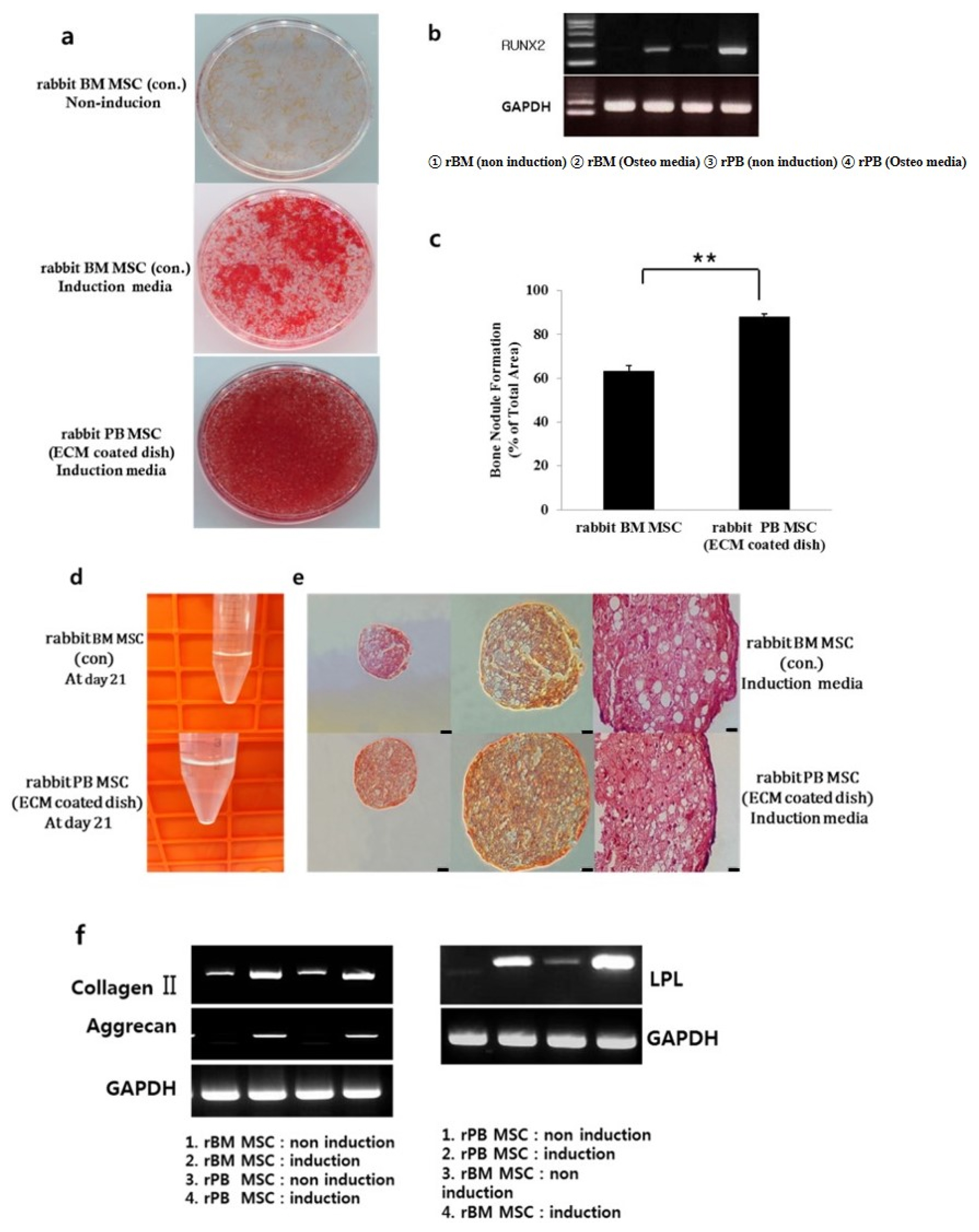
Fig. 4 Osteogenic and Chondrogenic differentiation of rabbit BM MSCs and rabbit PB MSCs in vitro. (a) Calcium accumulation revealed by alizarin red S staining at ×200 magnification. (b) Expression of RUNX-2, which induces osteoblast differentiation, in both experimental groups. (c) Percentage of mineralized area/total area of the dish. Data show mean±SE of three dishes (*p<0.05) (d) Aggregate pellet culture. Aliquots of rabbit BM MSCs and rabbit PB MSCs form a spherical pellet in 21 days. (e) Histologic sections were stained with Safranin-O. The stained images are presented as a whole sample (first columns, ×100) and at high magnification (second and last columns, ×200, ×400). (f) Total RNA was isolated from rabbit BM MSCs and rabbit PB MSCs and expression levels of type II collagen, aggrecan, and GAPDH were examined by RT-PCR analysis.
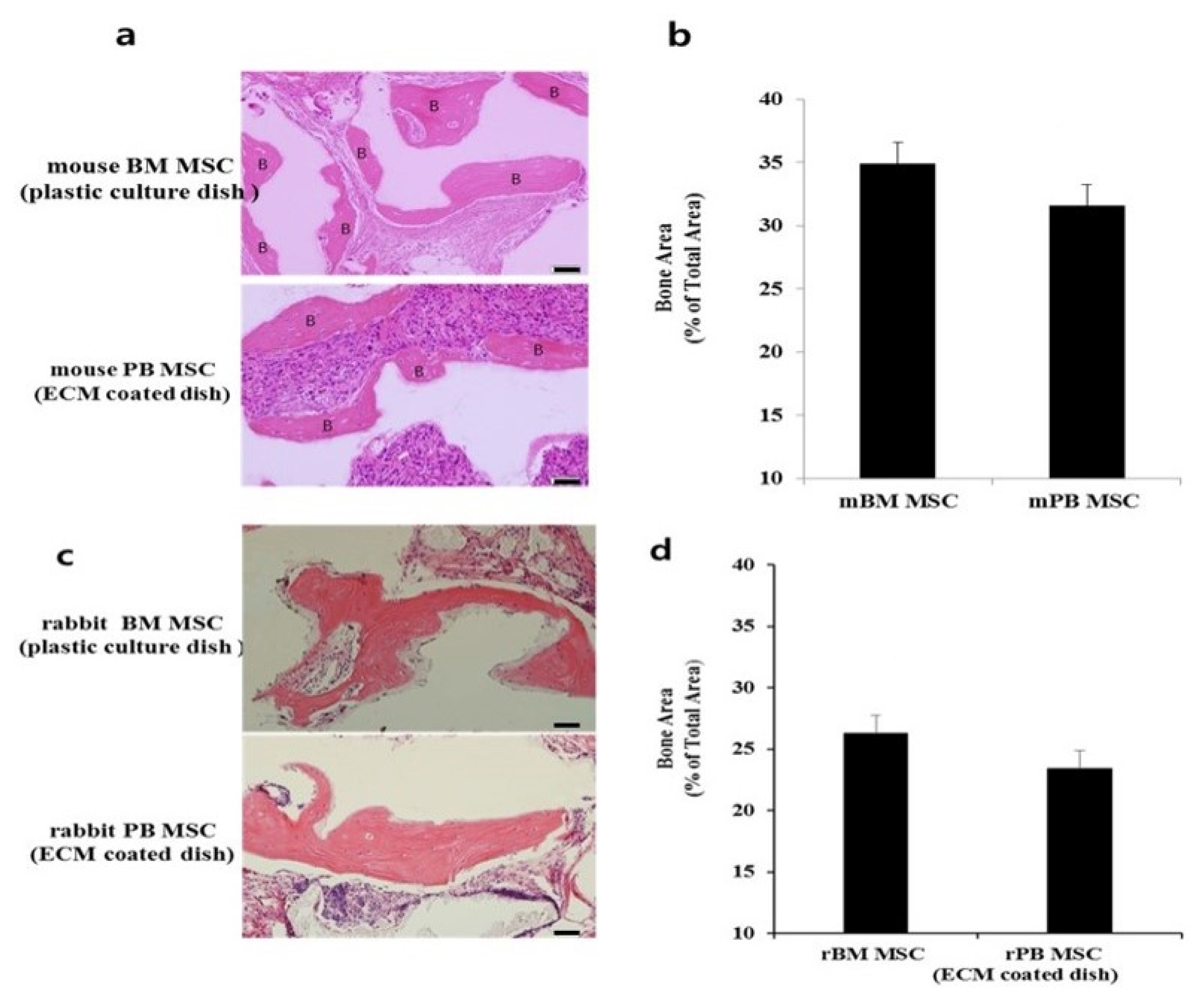
Fig. 5 Bone formation of BM MSCs and PB MSCs in vivo after transplantation into immunocompromised mice. (a, c) Bone formation by in vivo transplantation of mouse and rabbit MSCs. After 8 weeks, the transplants were harvested and sections were stained with H&E to evaluate bone formation. Scale bar=50 μm. (b, d) PB MSCs generate as much bone area as BM MSCs (*p>0.05). Data show mean±SE of bone area.
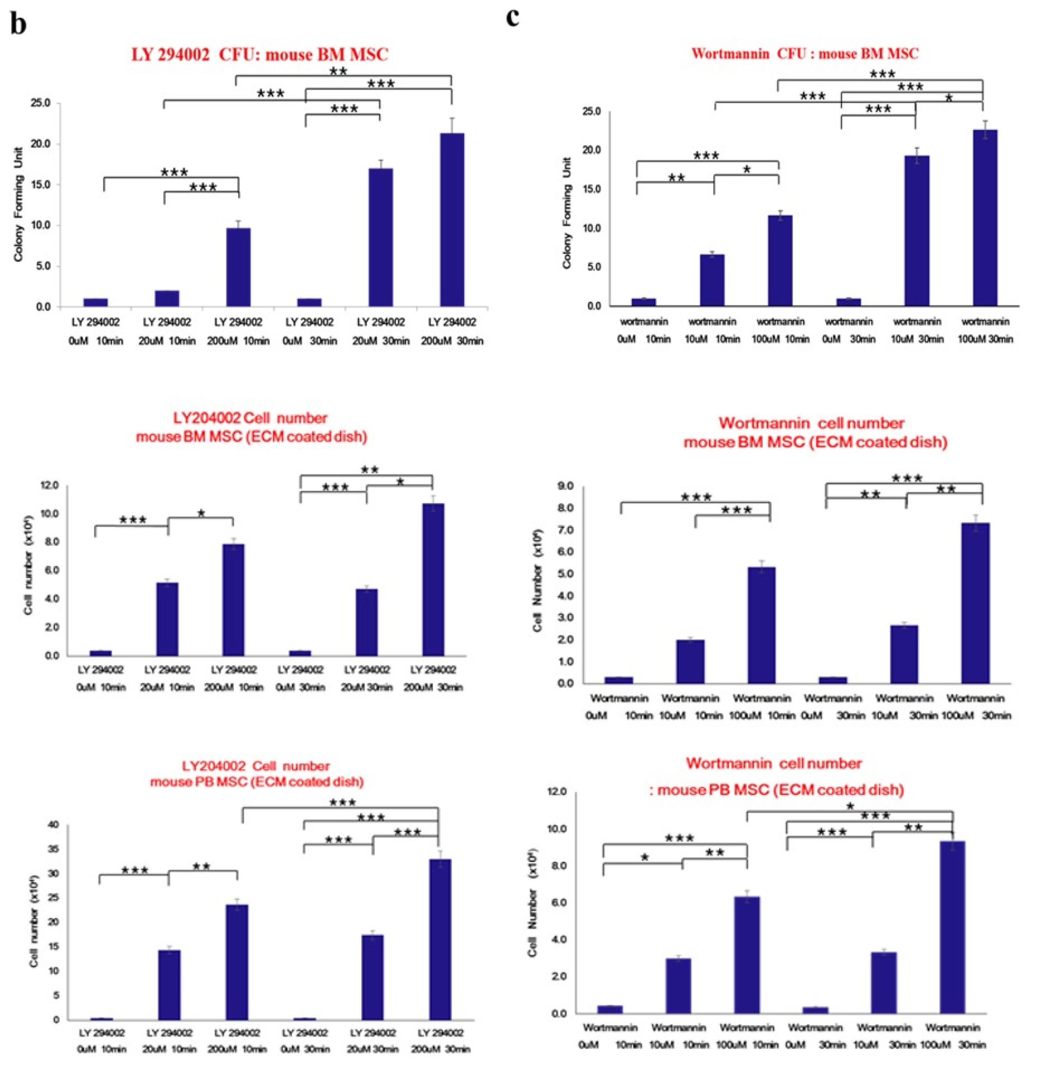
Fig. 6 PI3K modulates the number of adherent colony-forming unit-fibroblasts (CFU-Fs) in primary bone marrow cultures. (a) Hypothetical model showing the culture of mouse BM MSCs on culture dish for 16 days, before treatment with the PI3K inhibitors LY294002 (20 μM and 200 μM) and Wortmannin (10 μM and 100 μM) for 10 min and 30 min. The colony forming efficiency of cells from suspension was determined. (b, c) Mouse BM MSCs, mouse BM matrix cells, and mouse PB cells were treated with inhibitors at various concentrations and cell number was determined by trypan blue assay. Left panel: Colony forming assay of mouse BM MSCs in suspension after culture for 16 days and treatment with LY294002. Right panel: Colony forming assay of mouse BM MSCs in suspension after culture for 16 days and treatment with Wortmannin. Wortmannin treatment increased the number of cells in the supernatant in a dose- and time-dependent manner. Data are presented as means±SD. Statistical analysis was performed using Student’s t-tests. *Indicates significant difference versus control (*p<0.05; **p<0.01; ***p<0.001 by Student’s t-test).
Reference
-
References
1. Ullah I, Subbarao RB, Rho GJ. Human mesenchymal stem cells - current trends and future prospective. Biosci Rep. 2015; 35:pii: e00191. DOI: 10.1042/BSR20150025. PMID: 25797907. PMCID: 4413017.
Article2. Perlin JR, Sporrij A, Zon LI. Blood on the tracks: hematopoietic stem cell-endothelial cell interactions in homing and engraftment. J Mol Med (Berl). 2017; 95:809–819. DOI: 10.1007/s00109-017-1559-8. PMID: 28702683. PMCID: PMC5558790.
Article3. Xie C, Yang Z, Suo Y, Chen Q, Wei D, Weng X, Gu Z, Wei X. Systemically infused mesenchymal stem cells show different homing profiles in healthy and tumor mouse models. Stem Cells Transl Med. 2017; 6:1120–1131. DOI: 10.1002/sctm.16-0204. PMID: 28205428. PMCID: 5442841.
Article4. Chamberlain G, Fox J, Ashton B, Middleton J. Concise review: mesenchymal stem cells: their phenotype, differentiation capacity, immunological features, and potential for homing. Stem Cells. 2007; 25:2739–2749. DOI: 10.1634/stemcells.2007-0197. PMID: 17656645.
Article5. Trounson A, McDonald C. Stem cell therapies in clinical trials: progress and challenges. Cell Stem Cell. 2015; 17:11–22. DOI: 10.1016/j.stem.2015.06.007. PMID: 26140604.
Article6. Goldberg A, Mitchell K, Soans J, Kim L, Zaidi R. The use of mesenchymal stem cells for cartilage repair and regeneration: a systematic review. J Orthop Surg Res. 2017; 12:39. DOI: 10.1186/s13018-017-0534-y. PMID: 28279182. PMCID: 5345159.
Article7. Liotta F, Annunziato F, Castellani S, Boddi M, Alterini B, Castellini G, Mazzanti B, Cosmi L, Acquafresca M, Bartalesi F, Dilaghi B, Dorigo W, Graziani G, Bartolozzi B, Bellandi G, Carli G, Bartoloni A, Fargion A, Fassio F, Fontanari P, Landini G, Lucente EAM, Michelagnoli S, Orsi Battaglini C, Panigada G, Pigozzi C, Querci V, Santarlasci V, Parronchi P, Troisi N, Baggiore C, Romagnani P, Mannucci E, Saccardi R, Pratesi C, Gensini G, Romagnani S, Maggi E. Therapeutic efficacy of autologous non-mobilized enriched circulating endothelial progenitors in patients with critical limb ischemia - the SCELTA trial. Circ J. 2018; 82:1688–1698. DOI: 10.1253/circj.CJ-17-0720. PMID: 29576595.
Article8. Freitag J, Bates D, Boyd R, Shah K, Barnard A, Huguenin L, Tenen A. Mesenchymal stem cell therapy in the treatment of osteoarthritis: reparative pathways, safety and efficacy - a review. BMC Musculoskelet Disord. 2016; 17:230. DOI: 10.1186/s12891-016-1085-9. PMID: 27229856. PMCID: 4880954.
Article9. Zheng RC, Park YK, Cho JJ, Kim SK, Heo SJ, Koak JY, Lee JH. Bone regeneration at dental implant sites with suspended stem cells. J Dent Res. 2014; 93:1005–1013. DOI: 10.1177/0022034514548706. PMID: 25183420. PMCID: 4293714.
Article10. Romanov YA, Svintsitskaya VA, Smirnov VN. Searching for alternative sources of postnatal human mesenchymal stem cells: candidate MSC-like cells from umbilical cord. Stem Cells. 2003; 21:105–110. DOI: 10.1634/stemcells.21-1-105. PMID: 12529557.
Article11. Fahy N, Alini M, Stoddart MJ. Mechanical stimulation of mesenchymal stem cells: implications for cartilage tissue engineering. J Orthop Res. 2018; 36:52–63. DOI: 10.1002/jor.23670. PMID: 28763118.
Article12. Fadini GP, Ciciliot S, Albiero M. Concise review: perspectives and clinical implications of bone marrow and circulating stem cell defects in diabetes. Stem Cells. 2017; 35:106–116. DOI: 10.1002/stem.2445. PMID: 27401837.
Article13. Pineault N, Abu-Khader A. Advances in umbilical cord blood stem cell expansion and clinical translation. Exp Hematol. 2015; 43:498–513. DOI: 10.1016/j.exphem.2015.04.011. PMID: 25970610.
Article14. Zheng RC, Park YK, Kim SK, Cho J, Heo SJ, Koak JY, Lee SJ, Park JM, Lee JH, Kim JH. Bone regeneration of blood-derived stem cells within dental implants. J Dent Res. 2015; 94:1318–1325. DOI: 10.1177/0022034515590368. PMID: 26078421.
Article15. Wexler SA, Donaldson C, Denning-Kendall P, Rice C, Bradley B, Hows JM. Adult bone marrow is a rich source of human mesenchymal ‘stem’ cells but umbilical cord and mobilized adult blood are not. Br J Haematol. 2003; 121:368–374. DOI: 10.1046/j.1365-2141.2003.04284.x. PMID: 12694261.
Article16. Hassan G, Kasem I, Soukkarieh C, Aljamali M. A simple method to isolate and expand human umbilical cord derived mesenchymal stem cells: using explant method and umbilical cord blood serum. Int J Stem Cells. 2017; 10:184–192. DOI: 10.15283/ijsc17028. PMID: 28844128. PMCID: 5741200.
Article17. Ward PS, Thompson CB. Signaling in control of cell growth and metabolism. Cold Spring Harb Perspect Biol. 2012; 4:a006783. DOI: 10.1101/cshperspect.a006783. PMID: 22687276. PMCID: 3385956.
Article18. Scanlon V, Soung do Y, Adapala NS, Morgan E, Hansen MF, Drissi H, Sanjay A. Role of Cbl-PI3K interaction during skeletal remodeling in a murine model of bone repair. PLoS One. 2015; 10:e0138194. DOI: 10.1371/journal.pone.0138194. PMID: 26393915. PMCID: 4578922.
Article19. Fruman DA, Chiu H, Hopkins BD, Bagrodia S, Cantley LC, Abraham RT. The PI3K pathway in human disease. Cell. 2017; 170:605–635. DOI: 10.1016/j.cell.2017.07.029. PMID: 28802037. PMCID: 5726441.
Article20. Huang CY, Hagar KL, Frost LE, Sun Y, Cheung HS. Effects of cyclic compressive loading on chondrogenesis of rabbit bone-marrow derived mesenchymal stem cells. Stem Cells. 2004; 22:313–323. DOI: 10.1634/stemcells.22-3-313. PMID: 15153608.
Article21. Hidalgo-Bastida LA, Cartmell SH. Mesenchymal stem cells, osteoblasts and extracellular matrix proteins: enhancing cell adhesion and differentiation for bone tissue engineering. Tissue Eng Part B Rev. 2010; 16:405–412. DOI: 10.1089/ten.teb.2009.0714. PMID: 20163206.
Article22. Tondreau T, Meuleman N, Delforge A, Dejeneffe M, Leroy R, Massy M, Mortier C, Bron D, Lagneaux L. Mesenchymal stem cells derived from CD133-positive cells in mobilized peripheral blood and cord blood: proliferation, Oct4 expression, and plasticity. Stem Cells. 2005; 23:1105–1112. DOI: 10.1634/stemcells.2004-0330. PMID: 15955825.
Article23. He Q, Wan C, Li G. Concise review: multipotent mesenchymal stromal cells in blood. Stem Cells. 2007; 25:69–77. DOI: 10.1634/stemcells.2006-0335. PMID: 16973831.
Article24. Khosla S, Westendorf JJ, Mödder UI. Concise review: insights from normal bone remodeling and stem cell-based therapies for bone repair. Stem Cells. 2010; 28:2124–2128. DOI: 10.1002/stem.546. PMID: 20960512. PMCID: 3125598.
Article25. Wu G, Pan M, Wang X, Wen J, Cao S, Li Z, Li Y, Qian C, Liu Z, Wu W, Zhu L, Guo J. Osteogenesis of peripheral blood mesenchymal stem cells in self assembling peptide nanofiber for healing critical size calvarial bony defect. Sci Rep. 2015; 5:16681. DOI: 10.1038/srep16681. PMID: 26568114. PMCID: 4645224.
Article26. Lakkakorpi PT, Wesolowski G, Zimolo Z, Rodan GA, Rodan SB. Phosphatidylinositol 3-kinase association with the osteoclast cytoskeleton, and its involvement in osteoclast attachment and spreading. Exp Cell Res. 1997; 237:296–306. DOI: 10.1006/excr.1997.3797. PMID: 9434625.
Article27. Kim NG, Gumbiner BM. Adhesion to fibronectin regulates Hippo signaling via the FAK-Src-PI3K pathway. J Cell Biol. 2015; 210:503–515. DOI: 10.1083/jcb.201501025. PMID: 26216901. PMCID: 4523609.
Article28. Chen XD, Dusevich V, Feng JQ, Manolagas SC, Jilka RL. Extracellular matrix made by bone marrow cells facilitates expansion of marrow-derived mesenchymal progenitor cells and prevents their differentiation into osteoblasts. J Bone Miner Res. 2007; 22:1943–1956. DOI: 10.1359/jbmr.070725. PMID: 17680726.
Article
- Full Text Links
-
- Actions
-
Cited
- CITED
-
- Close
- Share
-
- Similar articles
-
- Concise Review: Differentiation of Human Adult Stem Cells Into Hepatocyte-like Cells In vitro
- The Role of Histone Acetylation in Mesenchymal Stem Cell Differentiation
- In vitro neuronal and osteogenic differentiation of mesenchymal stem cells from human umbilical cord blood
- Comparative Evaluation for Potential Differentiation of Endothelial Progenitor Cells and Mesenchymal Stem Cells into Endothelial-Like Cells
- Differential Potential of Stem Cells Following Their Origin: Subacromial Bursa, Bone Marrow, Umbilical Cord Blood
