Regenerative Medicine of the Bile Duct: Beyond the Myth
- Affiliations
-
- 1Department of Translational Medicine, Graduate School of Biomedical Science and Engineering, Hanyang University, Seoul, Korea. crane87@hanyang.ac.kr
- 2Department of Surgery, Hanyang University College of Medicine, Hanyang University, Seoul, Korea. thicknyh@gmail.com
- 3HY Indang Center of Regenerative Medicine and Stem Cell Research, Hanyang University, Seoul, Korea.
- KMID: 2465890
- DOI: http://doi.org/10.15283/ijsc18055
Abstract
- Cholangiopathies are rare diseases of the bile duct with high mortality rates. The current treatment for cholangiopathies is liver transplantation, but there are significant obstacles including a shortage of donors and a high risk of complications. Currently, there is only one available medicine on the market targeting cholangiopathies, and the results have been inadequate in clinical therapy. To overcome these obstacles, many researchers have used human induced pluripotent stem cells (hPSC) as a source for cholangiocyte-like cell generation and have incorporated advances in bioprinting to create artificial bile ducts for implantation and transplantation. This has allowed the field to move dramatically forward in studies of biliary regenerative medicine. In this review, the authors provide an overview of cholangiocytes, the organogenesis of the bile duct, cholangiopathies, and the current treatment and advances that have been made that are opening new doors to the study of cholangiopathies.
Keyword
MeSH Terms
Figure
-

Fig. 1 This figure and legend were adapted courtesy of the authors of Park et al. for use in this manuscript. (a) Photograph of the anastomosed bile duct. (b) The 3D-reconstructed MRCP image shows the anastomotic site and both the dilated intrahepatic bile ducts of the rabbit. (c) Photographs of the sectioned tissue containing the replaced artificial bile duct. Histopathological examination of the rabbit bile duct and liver. (d) Bile duct wall adjacent to the anastomosis site (black arrows). (e) Mucosal epithelium inside the bile duct wall showing normal regenerative atypia. (f) Liver parenchyma with well-preserved hepatic lobules and portal area.
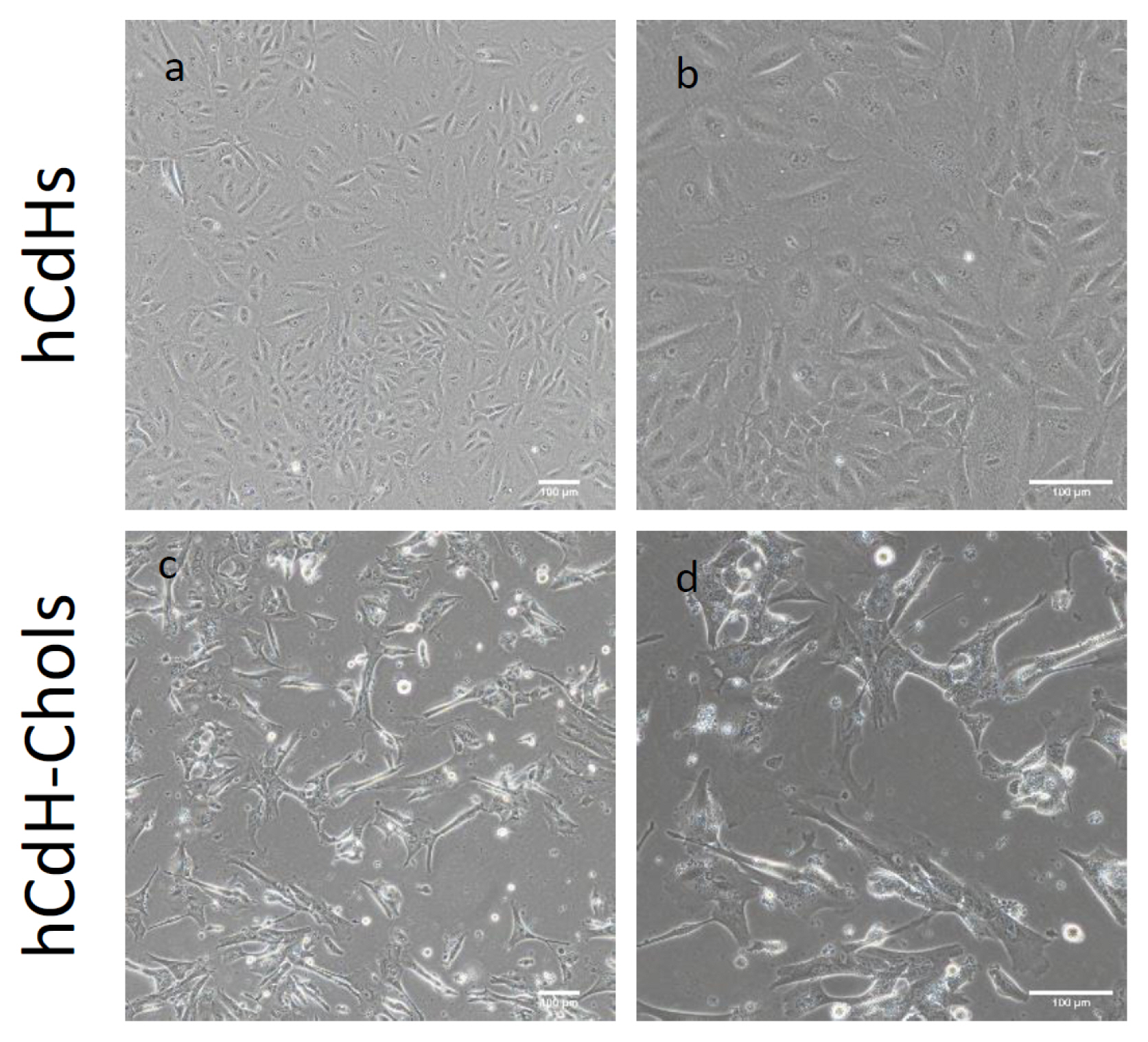
Fig. 2 Microscopy analysis of human CdH and human CdH derived cholangiocytes: (a) Morphological analysis of hCdH on a bright field microscope showing hCdH clusted together in a colony. (b) Magnified view of (a). Scale bars: 100 μm. (c) Bright field images showing hCdH-chol forming some branch-like structures similar to the cholangiocyte characteristics. (d) Magnified view of (c). Scale bars: 100 μm.

Fig. 3 Cholangiocyte markers of the relative expression between hCdH-chol and their negative control CdH as determined by RT-qpcr. GAPDH was used as the housekeeping gene. The data is shown as the mean value±SD. *p<.05, **p<.01, ***p<.001.
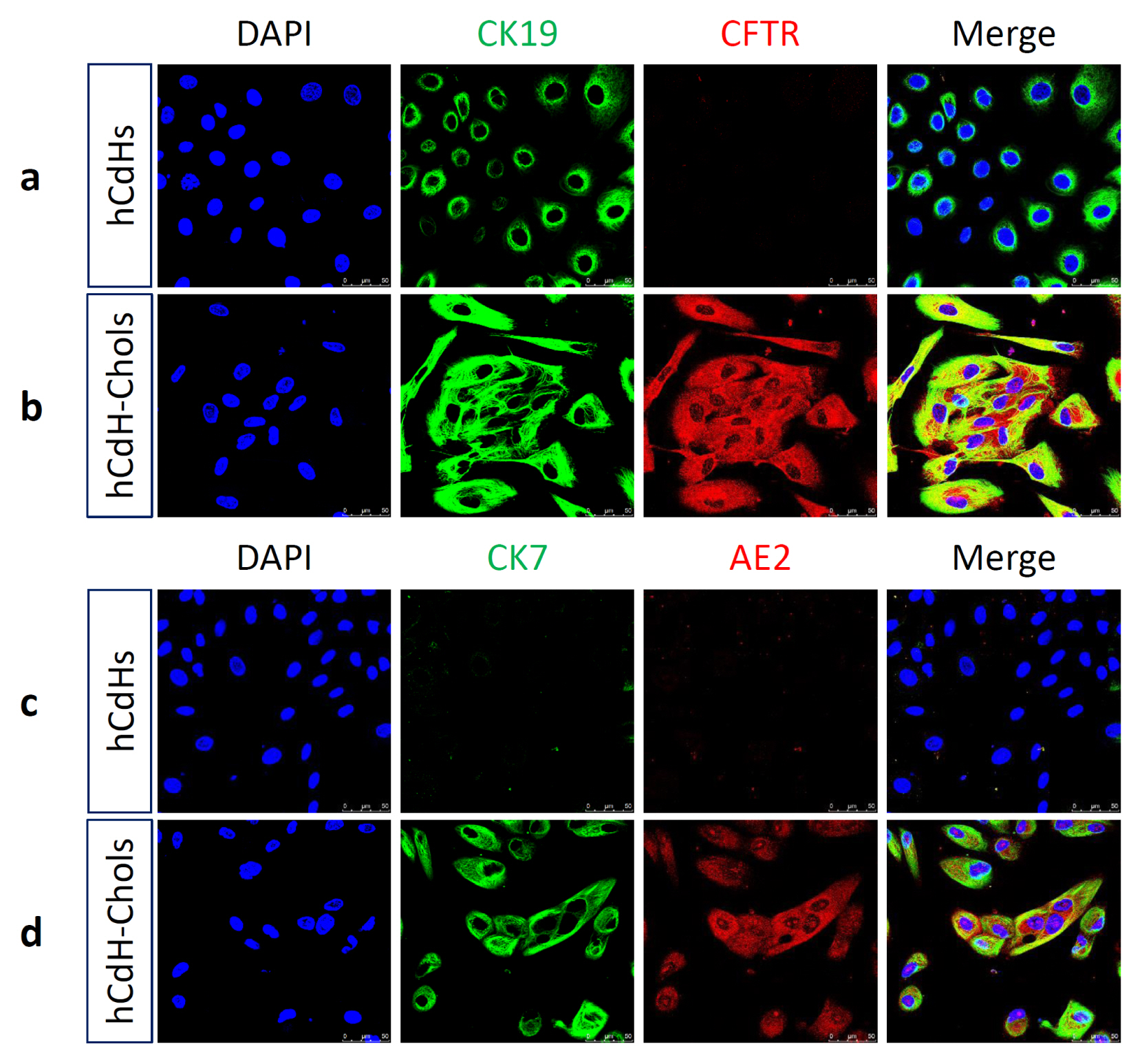
Fig. 4 hCdH-Chol expressed cholangiocyte markers seen through confocal microscopy. (a) Negative control hCdH stained with CK19 (green), CFTR (red). (b) CK19 (green), CFTR (red). (c) CK7 (green), AE2 (red). (d) CK7 (green), AE2 (red), and merged together showing a lack of expression of these genes. (d) hCdH-chol stained with CK7 (green) and AE2 (red) shows upregulation of these genes as seen through RT-qpcr. The nuclei were counterstained with Hoechst 33342 (blue). Scale bars, 50 μm.

Fig. 5 Fluorescein diacetate (FD) assay. (a) Brightfield images and fluorescence images were taken 0 min after FD removal showing uptake of FD by both hCdH and hCdH-chol. (b) Brightfield images and fluorescent images taken 30 minutes after FD removal show the functionality of hCdH-chols.
Reference
-
References
1. Tanimizu N, Miyajima A, Mostov KE. Liver progenitor cells develop cholangiocyte-type epithelial polarity in three-dimensional culture. Mol Biol Cell. 2007; 18:1472–1479. DOI: 10.1091/mbc.e06-09-0848. PMID: 17314404. PMCID: PMC1838984.
Article2. De Assuncao TM, Jalan-Sakrikar N, Huebert RC. Regenerative medicine and the biliary tree. Semin Liver Dis. 2017; 37:17–27. DOI: 10.1055/s-0036-1597818. PMID: 28201845. PMCID: PMC5479436.
Article3. O’Hara SP, Tabibian JH, Splinter PL, LaRusso NF. The dynamic biliary epithelia: molecules, pathways, and disease. J Hepatol. 2013; 58:575–582. DOI: 10.1016/j.jhep.2012.10.011. PMID: 23085249. PMCID: PMC3831345.
Article4. Cervantes-Alvarez E, Wang Y, Collin de l’Hortet A, Guzman-Lepe J, Zhu J, Takeishi K. Current strategies to generate mature human induced pluripotent stem cells derived cholangiocytes and future applications. Organogenesis. 2017; 13:1–15. DOI: 10.1080/15476278.2016.1278133. PMID: 28055309. PMCID: PMC5323032.
Article5. Tremblay KD, Zaret KS. Distinct populations of endoderm cells converge to generate the embryonic liver bud and ventral foregut tissues. Dev Biol. 2005; 280:87–99. DOI: 10.1016/j.ydbio.2005.01.003. PMID: 15766750.
Article6. Tabibian JH, Masyuk AI, Masyuk TV, O’Hara SP, LaRusso NF. Physiology of cholangiocytes. Compr Physiol. 2013; 3:541–565. PMID: 23720296. PMCID: PMC3831353.
Article7. Strazzabosco M, Fabris L. Development of the bile ducts: essentials for the clinical hepatologist. J Hepatol. 2012; 56:1159–1170. DOI: 10.1016/j.jhep.2011.09.022. PMID: 22245898. PMCID: PMC3328609.
Article8. Geisler F, Nagl F, Mazur PK, Lee M, Zimber-Strobl U, Strobl LJ, Radtke F, Schmid RM, Siveke JT. Liver-specific inactivation of Notch2, but not Notch1, compromises intrahepatic bile duct development in mice. Hepatology. 2008; 48:607–616. DOI: 10.1002/hep.22381. PMID: 18666240.
Article9. Cardinale V, Wang Y, Carpino G, Mendel G, Alpini G, Gaudio E, Reid LM, Alvaro D. The biliary tree--a reservoir of multipotent stem cells. Nat Rev Gastroenterol Hepatol. 2012; 9:231–240. DOI: 10.1038/nrgastro.2012.23. PMID: 22371217.
Article10. Shin S, Walton G, Aoki R, Brondell K, Schug J, Fox A, Smirnova O, Dorrell C, Erker L, Chu AS, Wells RG, Grompe M, Greenbaum LE, Kaestner KH. Foxl1-Cre-marked adult hepatic progenitors have clonogenic and bilineage differentiation potential. Genes Dev. 2011; 25:1185–1192. DOI: 10.1101/gad.2027811. PMID: 21632825. PMCID: PMC3110956.
Article11. Margagliotti S, Clotman F, Pierreux CE, Beaudry JB, Jacquemin P, Rousseau GG, Lemaigre FP. The Onecut transcription factors HNF-6/OC-1 and OC-2 regulate early liver expansion by controlling hepatoblast migration. Dev Biol. 2007; 311:579–589. DOI: 10.1016/j.ydbio.2007.09.013. PMID: 17936262.
Article12. Tanimizu N, Miyajima A. Notch signaling controls hepatoblast differentiation by altering the expression of liver-enriched transcription factors. J Cell Sci. 2004; 117:3165–3174. DOI: 10.1242/jcs.01169. PMID: 15226394.
Article13. Tchorz JS, Kinter J, Muller M, Tornillo L, Heim MH, Bettler B. Notch2 signaling promotes biliary epithelial cell fate specification and tubulogenesis during bile duct development in mice. Hepatology. 2009; 50:871–879. DOI: 10.1002/hep.23048. PMID: 19551907.
Article14. Zong Y, Panikkar A, Xu J, Antoniou A, Raynaud P, Lemaigre F, Stanger BZ. Notch signaling controls liver development by regulating biliary differentiation. Development. 2009; 136:1727–1739. DOI: 10.1242/dev.029140. PMID: 19369401. PMCID: PMC2673761.
Article15. Raynaud P, Carpentier R, Antoniou A, Lemaigre FP. Biliary differentiation and bile duct morphogenesis in development and disease. Int J Biochem Cell Biol. 2011; 43:245–256. DOI: 10.1016/j.biocel.2009.07.020. PMID: 19735739.
Article16. Hussain SZ, Sneddon T, Tan X, Micsenyi A, Michalopoulos GK, Monga SP. Wnt impacts growth and differentiation in ex vivo liver development. Exp Cell Res. 2004; 292:157–169. DOI: 10.1016/j.yexcr.2003.08.020. PMID: 14720515.
Article17. Zong Y, Stanger BZ. Molecular mechanisms of liver and bile duct development. Wiley Interdiscip Rev Dev Biol. 2012; 1:643–655. DOI: 10.1002/wdev.47. PMID: 23799566.
Article18. Lazaridis KN, LaRusso NF. The Cholangiopathies. Mayo Clin Proc. 2015; 90:791–800. DOI: 10.1016/j.mayocp.2015.03.017. PMID: 25957621. PMCID: PMC4533104.
Article19. McDaniell R, Warthen DM, Sanchez-Lara PA, Pai A, Krantz ID, Piccoli DA, Spinner NB. NOTCH2 mutations cause Alagille syndrome, a heterogeneous disorder of the notch signaling pathway. Am J Hum Genet. 2006; 79:169–173. DOI: 10.1086/505332. PMID: 16773578. PMCID: PMC1474136.
Article20. Kim J, Yang B, Paik N, Choe YH, Paik YH. A case of Alagille syndrome presenting with chronic cholestasis in an adult. Clin Mol Hepatol. 2017; 23:260–264. DOI: 10.3350/cmh.2016.0057. PMID: 28683534. PMCID: PMC5628001.
Article21. Choe JY, Kim H. Intrahepatic cholangiocarcinoma with predominant ductal plate malformation pattern. Clin Mol Hepatol. 2014; 20:214–217. DOI: 10.3350/cmh.2014.20.2.214. PMID: 25032189. PMCID: PMC4099338.
Article22. Rizvi S, Gores GJ. Pathogenesis, diagnosis, and management of cholangiocarcinoma. Gastroenterology. 2013; 145:1215–1229. DOI: 10.1053/j.gastro.2013.10.013. PMID: 24140396. PMCID: PMC3862291.
Article23. Kim KA, Jeong SH. The diagnosis and treatment of primary biliary cirrhosis. Korean J Hepatol. 2011; 17:173–179. DOI: 10.3350/kjhep.2011.17.3.173. PMID: 22102382. PMCID: PMC3304651.
Article24. Verkade HJ, Bezerra JA, Davenport M, Schreiber RA, Mieli-Vergani G, Hulscher JB, Sokol RJ, Kelly DA, Ure B, Whitington PF, Samyn M, Petersen C. Biliary atresia and other cholestatic childhood diseases: advances and future challenges. J Hepatol. 2016; 65:631–642. DOI: 10.1016/j.jhep.2016.04.032. PMID: 27164551.
Article25. Lazaridis KN, Strazzabosco M, Larusso NF. The cholangiopathies: disorders of biliary epithelia. Gastroenterology. 2004; 127:1565–1577. DOI: 10.1053/j.gastro.2004.08.006. PMID: 15521023.
Article26. Halilbasic E, Fuchs C, Hofer H, Paumgartner G, Trauner M. Therapy of primary sclerosing cholangitis--today and tomorrow. Dig Dis. 2015; 33(Suppl 2):149–163. DOI: 10.1159/000440827. PMID: 26641242.
Article27. Mousa HS, Carbone M, Malinverno F, Ronca V, Gershwin ME, Invernizzi P. Novel therapeutics for primary biliary cholangitis: toward a disease-stage-based approach. Autoimmun Rev. 2016; 15:870–876. DOI: 10.1016/j.autrev.2016.07.003. PMID: 27393766.
Article28. Merino-Azpitarte M, Lozano E, Perugorria MJ, Esparza-Baquer A, Erice O, Santos-Laso A, O’Rourke CJ, Andersen JB, Jimenez-Aguero R, Lacasta A, D’Amato M, Briz Ó, Jalan-Sakrikar N, Huebert RC, Thelen KM, Gradilone SA, Aransay AM, Lavín JL, Fernández-Barrena MG, Matheu A, Marzioni M, Gores GJ, Bujanda L, Marin JJG, Banales JM. SOX17 regulates cholangiocyte differentiation and acts as a tumor suppressor in cholangiocarcinoma. J Hepatol. 2017; 67:72–83. DOI: 10.1016/j.jhep.2017.02.017. PMID: 28237397. PMCID: PMC5502751.
Article29. Sampaziotis F, de Brito MC, Madrigal P, Bertero A, Saeb-Parsy K, Soares FAC, Schrumpf E, Melum E, Karlsen TH, Bradley JA, Gelson WT, Davies S, Baker A, Kaser A, Alexander GJ, Hannan NRF, Vallier L. Cholangiocytes derived from human induced pluripotent stem cells for disease modeling and drug validation. Nat Biotechnol. 2015; 33:845–852. DOI: 10.1038/nbt.3275. PMID: 26167629. PMCID: PMC4768345.
Article30. Sampaziotis F, Justin AW, Tysoe OC, Sawiak S, Godfrey EM, Upponi SS, Gieseck RL 3rd, de Brito MC, Berntsen NL, Gomez-Vazquez MJ, Ortmann D, Yiangou L, Ross A, Bargehr J, Bertero A, Zonneveld MCF, Pedersen MT, Pawlowski M, Valestrand L, Madrigal P, Georgakopoulos N, Pirmadjid N, Skeldon GM, Casey J, Shu W, Materek PM, Snijders KE, Brown SE, Rimland CA, Simonic I, Davies SE, Jensen KB, Zilbauer M, Gelson WTH, Alexander GJ, Sinha S, Hannan NRF, Wynn TA, Karlsen TH, Melum E, Markaki AE, Saeb-Parsy K, Vallier L. Reconstruction of the mouse extrahepatic biliary tree using primary human extrahepatic cholangiocyte organoids. Nat Med. 2017; 23:954–963. DOI: 10.1038/nm.4360. PMID: 28671689.
Article31. Dianat N, Dubois-Pot-Schneider H, Steichen C, Desterke C, Leclerc P, Raveux A, Combettes L, Weber A, Corlu A, Dubart-Kupperschmitt A. Generation of functional cholangiocyte-like cells from human pluripotent stem cells and HepaRG cells. Hepatology. 2014; 60:700–714. DOI: 10.1002/hep.27165. PMID: 24715669. PMCID: PMC4315871.
Article32. Ogawa M, Ogawa S, Bear CE, Ahmadi S, Chin S, Li B, Grompe M, Keller G, Kamath BM, Ghanekar A. Directed differentiation of cholangiocytes from human pluripotent stem cells. Nat Biotechnol. 2015; 33:853–861. DOI: 10.1038/nbt.3294. PMID: 26167630.
Article33. De Assuncao TM, Sun Y, Jalan-Sakrikar N, Drinane MC, Huang BQ, Li Y, Davila JI, Wang R, O’Hara SP, Lomberk GA, Urrutia RA, Ikeda Y, Huebert RC. Development and characterization of human-induced pluripotent stem cell-derived cholangiocytes. Lab Invest. 2015; 95:1218. DOI: 10.1038/labinvest.2015.99. PMID: 26412498.
Article34. Kido T, Koui Y, Suzuki K, Kobayashi A, Miura Y, Chern EY, Tanaka M, Miyajima A. CPM Is a useful cell surface marker to isolate expandable bi-potential liver progenitor cells derived from human iPS cells. Stem Cell Reports. 2015; 5:508–515. DOI: 10.1016/j.stemcr.2015.08.008. PMID: 26365514. PMCID: PMC4624956.
Article35. Aikawa M, Miyazawa M, Okamoto K, Toshimitsu Y, Torii T, Okada K, Akimoto N, Ohtani Y, Koyama I, Yoshito I. A novel treatment for bile duct injury with a tissue-engineered bioabsorbable polymer patch. Surgery. 2010; 147:575–580. DOI: 10.1016/j.surg.2009.10.049. PMID: 20004452.
Article36. Perez Alonso AJ, Del Olmo Rivas C, Romero IM, Canizares Garcia FJ, Poyatos PT. Tissue-engineering repair of extrahepatic bile ducts. J Surg Res. 2013; 179:18–21. DOI: 10.1016/j.jss.2012.08.035. PMID: 23010513.
Article37. Miyazawa M, Torii T, Toshimitsu Y, Okada K, Koyama I, Ikada Y. A tissue-engineered artificial bile duct grown to resemble the native bile duct. Am J Transplant. 2005; 5:1541–1547. DOI: 10.1111/j.1600-6143.2005.00845.x. PMID: 15888066.
Article38. Park SH, Kang BK, Lee JE, Chun SW, Jang K, Kim YH, Jeong MA, Kim Y, Kang K, Lee NK, Choi D, Kim HJ. Design and fabrication of a thin-walled free-form scaffold on the basis of medical image data and a 3D printed template: its potential use in bile duct regeneration. Acs Appl Mater Interfaces. 2017; 9:12290–12298. DOI: 10.1021/acsami.7b00849. PMID: 28322040.
Article
