Regeneration of Neural Networks in Immature Teeth with Non-Vital Pulp Following a Novel Regenerative Procedure
- Affiliations
-
- 1Department of Oral Diagnostic Sciences, Faculty of Dentistry, King Abdulaziz University, Jeddah, Saudi Arabia. maha.f.mounir@gmail.com
- 2Faculty of Dentistry, Alexandria University, Alexadria, Egypt.
- 3Department of Oral Biology, Faculty of Dentistry, Damanhour University, Damanhour, Egypt.
- 4Department of Oral Biology, King Abdulaziz University, Faculty of Dentistry, Jeddah, Saudi Arabia.
- KMID: 2465881
- DOI: http://doi.org/10.15283/ijsc19026
Abstract
- BACKGROUND AND OBJECTIVES
Recombinant amelogenin protein (RAP) was reported to induce soft-tissue regeneration in canine infected endodontically treated permanent teeth with open apices. To characterize identities of the cells found in the RAP regenerated tissues compared to authentic pulp by identifying: 1) stem cells by their expression of Sox2; 2) nerve fibers by distribution of the axonal marker peripherin; 3) axons by their expression of calcitonin gene-related peptide (CGRP); 4) the presence of astrocytes expressing glial fibrillary acidic proteins (GFAP).
METHODS
A total of 240 open-apex root canals in dogs were used. After establishment of oral contamination to the pulp, the canals were cleaned, irrigated, and 120 canals filled with RAP, and the other 120 with calcium hydroxide.
RESULTS
After 1, 3, and 6 months, teeth were recovered for immune-detection of protein markers associated with native pulp tissues. Regenerated pulp and apical papilla of RAP group revealed an abundance of stem cells showing intense immunoreactivity to Sox2 antibody, immunoreactivity of peripherin mainly in the A-fibers of the odontoblast layer and immunoreactivity to CGRP fibers in the central pulp region indicative of C-fibres. GFAP immunoreactivity was observed near the odontoblastic, cell-rich regions and throughout the regenerated pulp.
CONCLUSIONS
RAP induces pulp regeneration following regenerative endodontic procedures with cells identity by gene expression demonstrating a distribution pattern similar to the authentic pulp innervation. A- and C-fibers, as well as GFAP specific to astrocytic differentiation, are recognized. The origin of the regenerated neural networks may be derived from the Sox2 identified stem cells within the apical papilla.
Keyword
MeSH Terms
-
Amelogenin
Animals
Astrocytes
Axons
Calcitonin
Calcitonin Gene-Related Peptide
Calcium Hydroxide
Dental Pulp Cavity
Dental Pulp Necrosis
Dogs
Gene Expression
Glial Fibrillary Acidic Protein
Nerve Fibers
Odontoblasts
Periapical Periodontitis
Regeneration*
Stem Cells
Tooth*
Amelogenin
Calcitonin
Calcitonin Gene-Related Peptide
Calcium Hydroxide
Glial Fibrillary Acidic Protein
Figure
-

Fig. 1 Premolar teeth at selected time intervals following treatment with recombinant amelogenin and calcium hydroxide. (A) Apical and mid root area of a premolar tooth 1-month post treatment with r-amelogenin showing regenerated cellular tissue inside canal consistent with pulp tissue (P), and apical papilla (AP) and dentin (D). H&E stain. Original magnification, ×100. (B) Apical and mid root area of a premolar tooth 1-month post treatment with r-amelogenin showing cells in the regenerated pulp tissue (P), apical papilla (AP) and cellular dentin (CD) Trichrome stain. Original magnification, ×100. (C) Apical area of premolar tooth 1-month post treatment with r-amelogenin showing regenerated regenerated pulp tissue (P) in the root canal, dentin (D) and apical papilla (AP). H&E stain. Original magnification, ×100. (D) Apical and mid-root area of premolar tooth 1-month post treatment with r-amelogenin showing intense to moderate immunoreactivity to the peripherin (PER) protein. Intense immunoreactivity is observed at the odontoblast layer and odontoblastic zone in the form of parallel conical arrays. Moderate to intense immunoreactivity in the central root canal and pulp camber. Strong immunoreactivity is seen in the apical papilla (AP). Dentin (D). Peripherin antibody. Original magnification, ×100. (E) Apical area of premolar tooth 1-month post treatment with r-amelogenin showing intense to moderate immunoreactivity to the CGRP antigen in the cells of the apical papilla (AP) running axially in the central area of root canal (red lines). Dentin (D). CGRP antibody. Original magnification, ×100. (F) Apical area of premolar tooth 1-month post treatment with r-amelogenin showing intense immunoreactivity to the GFAP antibody in the odontoblastic region (arrow) and mild to moderate immunoreactivity throughout the regenerated pulp tissue. Mild immunoreactivity is seen in the cells of the apical papilla (AP). Dentin (D). GFAP antibody. Original magnification, ×40. (G) Apical area of premolar tooth 3-month post treatment with r-amelogenin showing intense immunoreactivity to peripherin antibody in the regenerated pulp tissue (P) that lies at the pulp periphery near the odontoblast area (red lines). Dentin (D), periodontal ligament PDL (black arrow). Peripherin antibody. Original magnification, ×40. (H) Apical area of premolar tooth 3-month post treatment with r-amelogenin showing moderate to intense immunoreactivity to CGRP antibody in the regenerated pulp tissue (P) that lies at root canal apex and in mid root in the central pulp region (red lines). Apical papilla (AP) shows moderate immunoreactivity. Dentin (D). CGRP antibody. Original magnification, ×100. (I) Apical area of premolar tooth 3-month post treatment with r-amelogenin showing moderate to intense immunoreactivity to GFAP antibody in the odontoblastic region (red arrow) and mild to moderate immunoreactivity throughout the regenerated pulp tissue (P). Dentin (D), periodontal ligament PDL (black arrow). GFAP antibody. Original magnification, ×100. (J) Apical area of premolar tooth 6-month post treatment with r-amelogenin showing intense immunoreactivity to peripherin antibody in the regenerated pulp tissue (P) that lies at pulp periphery near the odontoblast area (red lines). Dentin (D), PDL (black arrow). Peripherin antibody. Original magnification, ×100. (K) Apical area of premolar tooth 6-month post treatment with r-amelogenin showing intense immunoreactivity to CGRP antibody in the regenerated pulp tissue (P) that lies at root canal apex and in mid root in the central pulp region (red lines). Dentin (D), PDL (black arrow). CGRP antibody. Original magnification, ×100. (L) Apical and mid area of premolar tooth 6-month post treatment with r-amelogenin showing moderate to intense immunoreactivity to GFAP antibody in the odontoblastic and cell-rich zone regions (red arrows) and mild to moderate immunoreactivity throughout the regenerated pulp tissue (P). Dentin (D), PDL (black arrow). GFAP antibody. Original magnification, ×100. (M) Apical area of premolar tooth 1-month post treatment with calcium hydroxide showing no immunoreactivity to Sox2 antibody in the empty root canals (RC). Dentin (D). Mineralized island can be seen in the empty RC (box). Sox2 antibody. Original magnification, ×100. (N) Apical and mid root area of a premolar tooth 3-month post treatment with calcium hydroxide showing empty root canals (RC). Odontoblast cell shows intense immunereactivity to peripherin antibody. Dentin (D). Granulation tissue (GT) shows mild immune reactivity to peripherin antibody. Peripherin antibody. Original magnification, ×100. (O) Apical area of premolar tooth 6-month post treatment with calcium hydroxide showing no immunoreactivity to CGRP antibody in the empty root canals (RC). Dentin (D). Granulation tissue shows no immunereactivity to CGRP antibody. CGRP antibody. Original magnification, ×100.

Fig. 2 Premolar teeth at 1-month time interval following treatment with recombinant amelogenin. (A) Higher magnification of box in Fig. 1D showing positive peripherin Ag/Ab immunostaining (brown DAB chromogen) in 1~9 of 13 of the plotted areas, while 10~13 of the plotted areas show negative peripherin Ab/Ab reactivity (grey scale). Image analysis. Peripherin antibody. Original magnification, ×400. (B) Higher magnification of Fig. 1F showing positive GFAP Ag/Ab immunostaining (brown DAB chromogen) were detected in 1~20 out of 22 plotted areas. In contrast, 21~22 of the plotted areas showed negative GFAP Ab/Ab reactivity (grey scale). Image analysis. GFAP antibody. Original magnification, ×400.
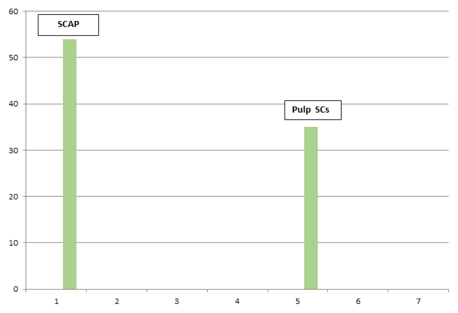
Fig. 3 Mean count for stem cells apical papilla SCAP+pulp stem cells. Quantitative image analysis immunohistochemical sections comparing between mean counts for SCAP+pulp stem cells (1000 pixel image scale/Mic. Mag. ×400).
Reference
-
References
1. Chai Y, Jiang X, Ito Y, Bringas P Jr, Han J, Rowitch DH, Soriano P, McMahon AP, Sucov HM. Fate of the mammalian cranial neural crest during tooth and mandibular morphogenesis. Development. 2000; 127:1671–1679. PMID: 10725243.
Article2. Fried K, Gibbs JL. Dental pulp innervation 6. Goldberg M, editor. The Dental Pulp: Biology, Pathology, and Regenerative Therapies. Berlin Heidelberg: Springer;2014. p. 75–95.3. Chandki R, Kala M, Banthia P, Banthia R. From stem to roots: Tissue engineering in endodontics. J Clin Exp Dent. 2012; 4:e66–e71. DOI: 10.4317/jced.50678. PMID: 24558528. PMCID: PMC3908813.
Article4. Yamauchi N, Nagaoka H, Yamauchi S, Teixeira FB, Miguez P, Yamauchi M. Immunohistological characterization of newly formed tissues after regenerative procedure in immature dog teeth. J Endod. 2011; 37:1636–1641. DOI: 10.1016/j.joen.2011.08.025. PMID: 22099896.
Article5. Lin LM, Di Fiore PM, Lin J, Rosenberg PA. Histological study of periradicular tissue responses to uninfected and infected devitalized pulps in dogs. J Endod. 2006; 32:34–38. DOI: 10.1016/j.joen.2005.10.010. PMID: 16410065.
Article6. Sharma S, Sikri V, Sharma NK, Sharma VM. Regenration of tooth pulp and dentin: trends and advances. Ann Neurosci. 2010; 17:31–43. DOI: 10.5214/ans.0972.7531.2010.170109.7. Kim TH, Bae YC, Yang ES. Distribution of peripherin immunoreactive axons in rat molar pulp. Korean J Phys Anthropol. 2013; 26:33–40. DOI: 10.11637/kjpa.2013.26.1.33.
Article8. Magloire H, Maurin JC, Couble ML, Shibukawa Y, Tsumura M, Thivichon-Prince B, Bleicher F. Topical review. Dental pain and odontoblasts: facts and hypotheses. J Orofac Pain. 2010; 24:335–349. PMID: 21197505.9. Uddman R, Grunditz T, Sundler F. Calcitonin gene related peptide: a sensory transmitter in dental pulps? Scand J Dent Res. 1986; 94:219–224. PMID: 3488582.
Article10. Veerayutthwilai O, Luis NA, Crumpton RM, MacDonald GH, Byers MR. Peripherin- and CGRP-immunoreactive nerve fibers in rat molars have different locations and developmental timing. Arch Oral Biol. 2006; 51:748–760. DOI: 10.1016/j.archoralbio.2006.03.011. PMID: 16720017.
Article11. Kökten T, Lesot H, Kuchler-Bopp S. Experimental design for the innervation of tooth forming from implanted cell re-associations. Eberli D, editor. Cells and Biomaterials in Regenerative Medicine. Rijeka: IntechOpen;2014. p. 345–373.
Article12. Chen FM, Wu LA, Zhang M, Zhang R, Sun HH. Homing of endogenous stem/progenitor cells for in situ tissue regeneration: promises, strategies, and translational perspectives. Biomaterials. 2011; 32:3189–3209. DOI: 10.1016/j.biomaterials.2010.12.032. PMID: 21300401.
Article13. Yeo JC, Ng HH. The transcriptional regulation of pluripotency. Cell Res. 2013; 23:20–32. DOI: 10.1038/cr.2012.172. PMID: 23229513. PMCID: PMC3541660.
Article14. Zhang L, Yuan G, Liu H, Lin H, Wan C, Chen Z. Expression pattern of Sox2 during mouse tooth development. Gene Expr Patterns. 2012; 12:273–281. DOI: 10.1016/j.gep.2012.07.001. PMID: 22835638.
Article15. Zhang S, Cui W. Sox2, a key factor in the regulation of pluripotency and neural differentiation. World J Stem Cells. 2014; 6:305–311. DOI: 10.4252/wjsc.v6.i3.305. PMID: 25126380. PMCID: PMC4131272.
Article16. Farahani RM, Simonian M, Hunter N. Blueprint of an ancestral neurosensory organ revealed in glial networks in human dental pulp. J Comp Neurol. 2011; 519:3306–3326. DOI: 10.1002/cne.22701. PMID: 21681747.
Article17. Mounir MM, Matar MA, Lei Y, Snead ML. Recombinant amelogenin protein induces apical closure and pulp regeneration in open-apex, nonvital permanent canine teeth. J Endod. 2016; 42:402–412. DOI: 10.1016/j.joen.2015.11.003. PMID: 26709200. PMCID: PMC4766029.
Article18. Hargreaves KM, Diogenes A, Teixeira FB. Treatment options: biological basis of regenerative endodontic procedures. J Endod. 2013; 39(3 Suppl):S30–S43. DOI: 10.1016/j.joen.2012.11.025. PMID: 23439043. PMCID: PMC3589799.
Article19. Wang X, Thibodeau B, Trope M, Lin LM, Huang GT. Histologic characterization of regenerated tissues in canal space after the revitalization/revascularization procedure of immature dog teeth with apical periodontitis. J Endod. 2010; 36:56–63. DOI: 10.1016/j.joen.2009.09.039. PMID: 20003936.
Article20. Nakashima M, Akamine A. The application of tissue engineering to regeneration of pulp and dentin in endodontics. J Endod. 2005; 31:711–718. DOI: 10.1097/01.don.0000164138.49923.e5. PMID: 16186748.
Article21. Nör JE. Tooth regeneration in operative dentistry. Oper Dent. 2006; 31:633–642. DOI: 10.2341/06-000. PMID: 17153970.22. Smith AJ, Lumley PJ, Tomson PL, Cooper PR. Dental regeneration and materials: a partnership. Clin Oral Investig. 2008; 12:103–108. DOI: 10.1007/s00784-008-0189-5. PMID: 18270756.23. Simon S, Smith AJ. Regenerative endodontics. Br Dent J. 2014; 216:E13. DOI: 10.1038/sj.bdj.2014.243. PMID: 24651364.
Article24. Janjić K, Cvikl B, Moritz A, Agis H. Dental pulp regeneration: in vitro and in vivo approaches. Int J Stomatol Occlusion Med. 2016; 8:1–9. DOI: 10.1007/s12548-015-0139-1.25. Nakashima M, Iohara K. Regeneration of dental pulp by stem cells. Adv Dent Res. 2011; 23:313–319. DOI: 10.1177/0022034511405323. PMID: 21677085.
Article26. Yang J, Yuan G, Chen Z. Pulp regeneration: current approaches and future challenges. Front Physiol. 2016; 7:58. DOI: 10.3389/fphys.2016.00058. PMID: 27014076. PMCID: PMC4779938.
Article27. Amin HD, Olsen I, Knowles J, Dard M, Donos N. A tyrosine-rich amelogenin peptide promotes neovasculogenesis in vitro and ex vivo. Acta Biomater. 2014; 10:1930–1939. DOI: 10.1016/j.actbio.2013.11.027. PMID: 24321350.
Article28. Jonke E, Gemperli AC, Zhang T, Özdemir B, Dard M, Rausch-Fan X, Andrukhov O. Effect of tyrosine-rich amelogenin peptide on behavior and differentiation of endothelial cells. Clin Oral Investig. 2016; 20:2275–2284. DOI: 10.1007/s00784-016-1726-2. PMID: 26867593. PMCID: PMC5069334.
Article29. Wang Z, Oron E, Nelson B, Razis S, Ivanova N. Distinct lineage specification roles for NANOG, OCT4, and SOX2 in human embryonic stem cells. Cell Stem Cell. 2012; 10:440–454. DOI: 10.1016/j.stem.2012.02.016. PMID: 22482508.
Article30. Graham V, Khudyakov J, Ellis P, Pevny L. SOX2 functions to maintain neural progenitor identity. Neuron. 2003; 39:749–765. DOI: 10.1016/S0896-6273(03)00497-5. PMID: 12948443.
Article31. Mimeault M, Batra SK. Great promise of tissue-resident adult stem/progenitor cells in transplantation and cancer therapies. Adv Exp Med Biol. 2012; 741:171–186. DOI: 10.1007/978-1-4614-2098-9_12. PMID: 22457110. PMCID: PMC3645316.
Article
- Full Text Links
-
- Actions
-
Cited
- CITED
-
- Close
- Share
-
- Similar articles
-
- A review of the regenerative endodontic treatment procedure
- Pulp tissue regeneration and root formation of permanent teeth with pulpal/periapical deseases
- Regenerative Endodontic Procedure in Korean Children and Adolescents: A Case Report
- Regenerating the Pulp–Dentine Complex Using Autologous Platelet Concentrates: A Critical Appraisal of the Current Histological Evidence
- Clinical and radiographic outcomes of regenerative endodontic treatment performed by endodontic postgraduate students: a retrospective study
