Vascular Leiomyoma of the Nasal Floor: The Risk of Misdiagnosis
- Affiliations
-
- 1Department of Otorhinolaryngology-Head and Neck Surgery, Konyang University College of Medicine, Daejeon, Korea. ismi96@kyuh.ac.kr
- 2Department of Pathology, Konyang University College of Medicine, Daejeon, Korea.
- KMID: 2465547
- DOI: http://doi.org/10.18787/jr.2019.26.2.132
Abstract
- Vascular leiomyoma (VL) is a rare tumor, particularly in the sinonasal region. The etiology of VL is unclear and there are a number of theories regarding how VL develops. To date, approximately 20 cases of VL have been reported in the sinonasal area. This is the first reported case of VL in the right nasal floor. We present the case of an adult patient with mild nasal obstruction and discomfort. Preoperative examination failed to accurately diagnose the patient with VL. Transnasal endoscopic resection was performed. There were no complications or recurrence postoperatively.
Keyword
Figure
-

Fig. 1 Endoscopic finding of a soft tissue mass in the right nasal floor. The mass has a round, reddish, and well-vascularized appearance.
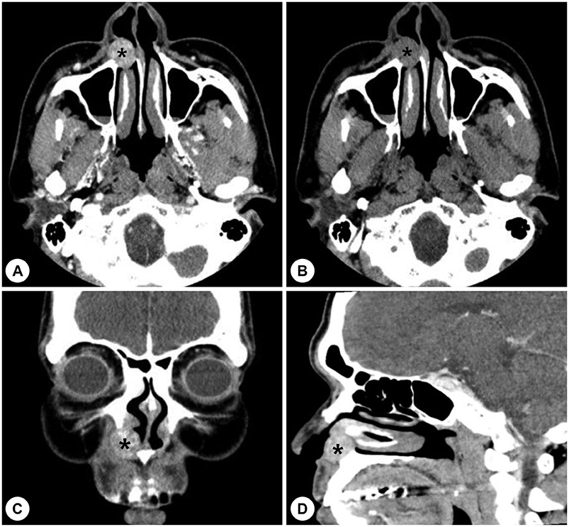
Fig. 2 Preoperative CT scan revealed a slightly heterogenously enhanced mass with bony remodeling in the maxilla. Black asterisk indicates soft tissue density lesion of the right nasal floor. A: enhanced axial. B: non-enhanced axial. C: enhanced coronal. D: enhanced sagittal cut.

Fig. 3 Preoperative MRI showing the soft tissue tumor in the right nasal floor. A: T1 imaging showing an iso-signal intensity, B: T2 imaging showing mixed low and high signal intensities. C: T1 Gd-enhanced imaging showing a slightly heterogenous enhancement.
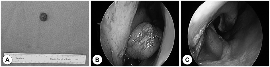
Fig. 4 The excised mass consists of a round, pinkish, and smooth soft tissue. Operative findings showed a well-capsulated soft tissue tumor in the right nasal floor without adhesion or bleeding. A: gross specimen. B: 30-degree endoscope view of the tumor removal. C: 0-degree endoscope view after tumor removal.
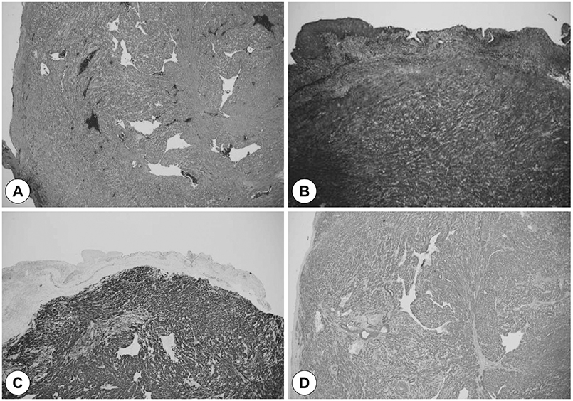
Fig. 5 Pathologic findings. Microscopically, the mass was composed of spindle cells with irregularly dilated, staghorn-shaped vessels. Masson's trichrome staining revealed fuchsinophilia of the spindle cells. Immunohistochemistry revealed a diffuse positive reaction for desmin and α-smooth muscle actin. A: Hematoxylin and eosin stain. B: Masson's trichrome stain. C: Immunohistochemistry for desmin. D: Immunohistochemistry for α-smooth muscle actin.
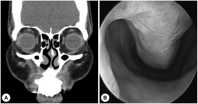
Fig. 6 Postoperative CT and endoscopic finding revealed no enhanced or residual mass in the right nasal floor. A: enhanced coronal. B: 0-degree endoscope.
Cited by 1 articles
-
Vascular Leiomyoma Arising From the Nasal Floor Misdiagnosed as a Nasolabial Cyst in an 74-Year-Old Patient
Jong Hwan Lee, Seung Yup Son, Kun Hee Lee
Korean J Otorhinolaryngol-Head Neck Surg. 2024;67(2):113-116. doi: 10.3342/kjorl-hns.2023.00353.
Reference
-
1. Duhig JT, Ayer JP. Vascular leiomyoma. A study of sixtyone cases. Arch Pathol. 1959; 68:424–430.2. Hachisuga T, Hashimoto H, Enjoji M. Angioleiomyoma. A clinicopathologic reappraisal of 562 cases. Cancer. 1984; 54(1):126–130.
Article3. Morimoto N. Angiomyoma (vascular leiomyoma): a clinico-pathologic study. Med J Kogoshima Univ. 1973; 24:663–683.4. Anderson TD, Weinstein GS. Recurrent angiomyoma (vascular leiomyoma) of the larynx after laser excision. Otolaryngol Head Neck Surg. 2000; 123(5):646–647.
Article5. Wang CP, Chang YL, Sheen TS. Vascular leiomyoma of the head and neck. Laryngoscope. 2004; 114(4):661–665.
Article6. Maesaka A, Keyaki Y, Nakahashi T. Nasal angioleiomyoma and leiomyosarcoma. Report of two cases. Otologia(Fukuoka). 1966; 12:42–47.7. Yoon TM, Yang HC, Choi YD, Lee DH, Lee JK, Lim SC. Vascular leiomyoma in the head and neck region: 11 years experience in one institution. Clin Exp Otorhinolaryngol. 2013; 6(3):171–175.
Article8. Liu Y, Li B, Li L, Liu Y, Wang C, Zha L. Angioleiomyomas in the head and neck: A retrospective clinical and immunohistochemical analysis. Oncol Lett. 2014; 8(1):241–247.
Article9. Agaimy A, Michal M, Thompson LD, Michal M. Angioleiomyoma of the Sinonasal Tract: Analysis of 16 Cases and Review of the Literature. Head Neck Pathol. 2015; 9(4):463–473.
Article10. Ramesh P, Annapureddy SR, Khan F, Sutaria PD. Angioleiomyoma: a clinical, pathological and radiological review. Int J Clin Pract. 2004; 58(6):587–591.
Article11. Brooks JK, Nikitakis NG, Goodman NJ, Levy BA. Clinicopathologic characterization of oral angioleiomyomas. Oral Surg Oral Med Oral Pathol Oral Radiol Endod. 2002; 94(2):221–227.
Article12. Gaitan Cepeda LA, Quezada Rivera D, Tenorio Rocha F, Leyva Huerta ER, Mendez Sanchez ER. Vascular leiomyoma of the oral cavity. Clinical, histopathological and immunohistochemical characteristics. Presentation of five cases and review of the literature. Med Oral Patol Oral Cir Bucal. 2008; 13(8):E483–E488.13. Maeda Y, Hirota J, Osaki T, Hayashi K, Sonobe H, Otsuki Y. Angiomyoma of the upper lip: report of a case with electron microscopic and immunohistochemical observation. Br J Oral Maxillofac Surg. 1989; 27(3):236–242.
Article14. Tseng PY, Lai YS, Chen MK, Shen KH. Progesterone receptor expression in sinonasal leiomyoma: a case report and review of the literature. Int J Clin Exp Pathol. 2014; 7(3):1224–1228.15. Vafiadis M, Kantas I, Panopoulou M, Sivridis E, Exarchakos G. Vascular leiomyoma of the nasal vestibule. Case report and literature review. B-ENT. 2008; 4(2):105–110.
- Full Text Links
-
- Actions
-
Cited
- CITED
-
- Close
- Share
-
- Similar articles
-
- Vascular Leiomyoma Arising From the Nasal Floor Misdiagnosed as a Nasolabial Cyst in an 74-Year-Old Patient
- A Case of Vascular Leiomyoma in Nasal Cavity: Case Report and Literature Review
- A Case of Vascular Leiomyoma of the Nasal Vestibule
- A Case of Leiomyoma of the Nasal Septum
- A Case of Angioleiomyoma Arising from the Right Inferior Turbinate
