Bronchogenic Cyst in Posterior Mediastinum with Butterfly Vertebra: A Case Report
- Affiliations
-
- 1Department of Radiology, St. Vincent's Hospital, College of Medicine, The Catholic University of Korea, Seoul, Korea. radiodoc@catholic.ac.kr
- 2Department of Pathology, St. Vincent's Hospital, College of Medicine, The Catholic University of Korea, Seoul, Korea.
- KMID: 2464928
- DOI: http://doi.org/10.3348/jksr.2019.80.6.1281
Abstract
- Most mediastinal cystic masses result from abnormal development of the embryo. Common developmental cysts in the posterior mediastinum are bronchogenic, neurenteric, and esophageal duplication cysts. These cystic masses appear identical on CT, but a cystic lesion adjacent to the esophagus is usually an esophageal duplication cyst, and a cyst associated with vertebral anomalies indicates a presumptive diagnosis of a neurenteric cyst. Herein, we present the case of a 27-year-old man with a bronchogenic cyst in the posterior mediastinum with a butterfly vertebra.
MeSH Terms
Figure
-
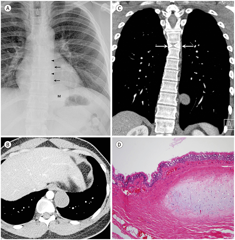
Fig. 1 Bronchogenic cyst in the posterior mediastinum with butterfly vertebra in a 27-year-old man. A. Chest X-ray image shows a well-defined mass (M) in the left paravertebral area, obscuring the left paraaortic interface (arrows) and paravertebral stripe (arrowheads). These findings suggest that the mass is located in the posterior mediastinum. The curvature of the thoracolumbar spines is S-shaped, representing mild scoliosis. B. Contrast-enhanced axial CT image shows a cystic mass with homogeneous fluid attenuation in the left paravertebral area, at the 11th thoracic vertebral level. The cyst wall has no definite boundary or calcifications. C. Coronal CT image shows that the 6th thoracic vertebral body has a sagittal cleft with a butterfly appearance (arrows). The cystic mass is seen partly caudal to the vertebral defect. Scoliosis is also seen. D. Micrograph shows ciliated pseudostratified columnar epithelium, smooth muscle fibers, and mature cartilage (hematoxylin and eosin stain, × 100).
Reference
-
1. Vargas D, Suby-Long T, Restrepo CS. Cystic lesions of the mediastinum. Semin Ultrasound CT MR. 2016; 37:212–222.2. Lee SH, Dante SJ, Simeone FA, Curtis MT. Thoracic neurenteric cyst in an adult: case report. Neurosurgery. 1999; 45:1239–1242. disscussion 1242–1243.3. Ranganath SH, Lee EY, Restrepo R, Eisenberg RL. Mediastinal masses in children. AJR Am J Roentgenol. 2012; 198:W197–W216.4. Strollo DC, Rosado-de-Christenson ML, Jett JR. Primary mediastinal tumors: part II. Tumors of the middle and posterior mediastinum. Chest. 1997; 112:1344–1357.5. Yoon JH, Lee KN, Kang EJ, Kim MS, Choi PJ, Roh MS. Bronchogenic cyst in an intradiaphragmatic location: a case report. J Korean Soc Radiol. 2018; 79:227–232.6. Kawashima A, Fishman EK, Kuhlman JE, Nixon MS. CT of posterior mediastinal masses. Radiographics. 1991; 11:1045–1067.7. Vilela TT, Daher RT, Nóbrega MD, Ximenes Filho CA, Montandon C, Montandon Júnior ME. Congenital mediastinal cysts: imaging findings. Radiologia Brasileira. 2009; 42:57–62.8. Occhipinti M, Heidinger BH, Franquet E, Eisenberg RL, Bankier AA. Imaging the posterior mediastinum: a multimodality approach. Diagn Interv Radiol. 2015; 21:293–306.
- Full Text Links
-
- Actions
-
Cited
- CITED
-
- Close
- Share
-
- Similar articles
-
- Intramural Bronchogenic Cyst of the Esophagus: A case report
- Retroperitoneal Bronchogenic Cyst Located in the Presacral Space: A Case Report
- A case of Bronchogenic Cyst in Diaphragm
- Multiple bronchogenic cysts: A case of multiple locations of mediastinum and lung parenchyme
- Mediastinal Bronchogenic Cyst Misdiagnosed as Asthma and Dysphagia in a Child: One Case Report
