Malignant Gastric Paraganglioma with Liver Metastasis: Imaging Findings and Literature Review
- Affiliations
-
- 1Department of Radiology, Wonkwang University Hospital, Wonkwang University College of Medicine, Iksan, Korea. yjyh@wku.ac.kr
- 2Department of Pathology, Wonkwang University Hospital, Wonkwang University College of Medicine, Iksan, Korea.
- KMID: 2464924
- DOI: http://doi.org/10.3348/jksr.2019.80.6.1258
Abstract
- Paraganglioma is a neuroendocrine tumor that grows in organs other than the adrenal gland and typically originates in the chromaffin cells, which are distributed along the regions of the sympathetic and parasympathetic branches of the nervous system in the entire body. Abdominal paraganglioma mainly arises in the retroperitoneum, which lines the sympathetic nerve branches; it rarely arises in other anatomical structures. In only a few cases reported in the literature, paraganglioma originated from the stomach. According to the current and previous case reports, gastric paraganglioma seems to be a hypervascular tumor growing around the stomach; it can be easily contrast-enhanced and may be accompanied by distant metastases.
MeSH Terms
Figure
-
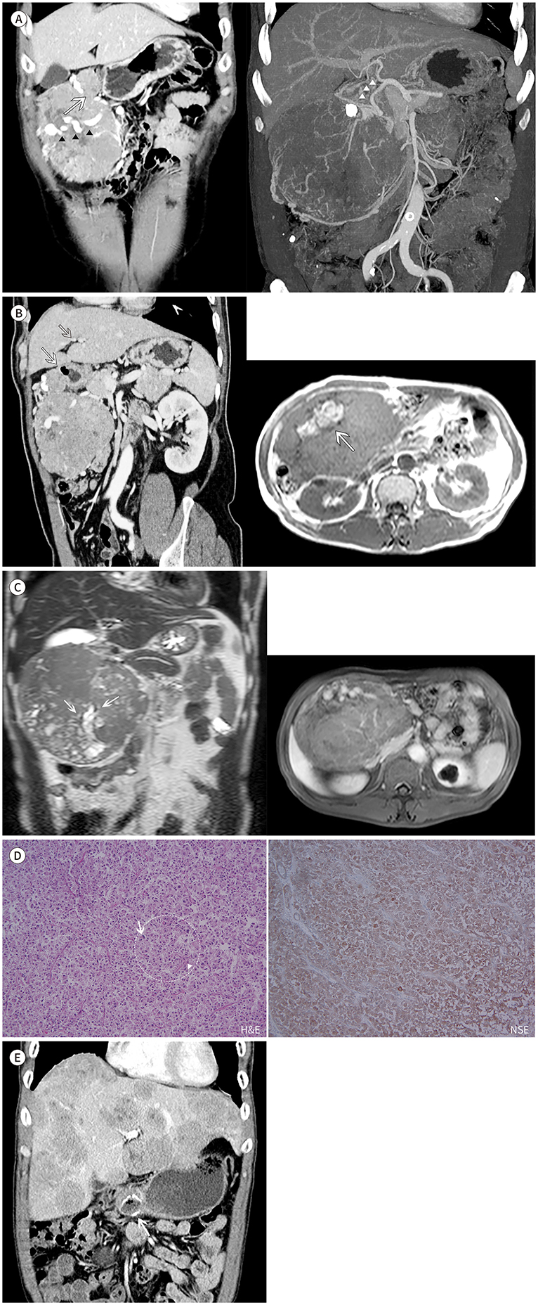
Fig. 1 Gastric paraganglioma in a 61-year-old man. A. Coronal portal-phase CT shows a large hypervascular mass, with heterogeneous enhancement in the right upper quadrant of the abdomen. The large mass from the gastric antrum (arrow) has a tortuous peritumoral and intratumoral vessels (arrowheads) (left image). Coronal maximal intensity projection CT reveals peritumoral and intratumoral vessels arising in the right gastric, gastroduodenal, gastroepiploic (arrowheads), and superior mesenteric arteries (right image). B. Multiplanar reformatted CT shows that the large mass arises from the gastric antrum (arrow) (left image). Axial T1-weighted in-phase MRI shows hyperintense lesions, which indicate hemorrhage (arrow) (right image). C. Coronal T2-weighted MRI reveals a large heterogeneous hyperintense mass with internal signal void vessels (arrows). Axial T1-weighted fat-suppressed contrast-enhanced MRI shows a heterogeneous enhancing mass with hypervascularity in the gastric antrum. D. Photomicrograph shows epithelioid (arrow) and mesenchymal cells (arrowhead) with some portion of nested pattern (dotted circle) (left, hematoxylin and eosin, × 100). Immunohistochemistry shows tumor cells positive for neuron-specific enolase (right, × 200). E. In the 1-year follow-up, axial portal-phase CT shows multiple enhancing liver masses, confirmed as metastases from the paraganglioma on liver biopsy. Surgical clips after mass excision from the gastric antrum are shown (arrow).
Reference
-
1. Lee KY, Oh YW, Noh HJ, Lee YJ, Yong HS, Kang EY, et al. Extraadrenal paragangliomas of the body: imaging features. AJR Am J Roentgenol. 2006; 187:492–504.2. Chrisoulidou A, Kaltsas G, Ilias I, Grossman AB. The diagnosis and management of malignant phaeochromocytoma and paraganglioma. Endocr Relat Cancer. 2007; 14:569–585.3. Westbrook KC, Bridger WM, Williams GD. Malignant nonchromaffin paraganglioma of the stomach. Am J Surg. 1972; 124:407–409.4. Schmid C, Beham A, Steindorfer P, Auböck L, Waltner F. Non-functional malignant paraganglioma of the stomach. Virchows Arch A Pathol Anat Histopathol. 1990; 417:261–266.5. Crosbie J, Humphreys WG, Maxwell M, Maxwell P, Cameron CH, Toner PG. Gastric paraganglioma: an immunohistological and ultrastructural case study. J Submicrosc Cytol Pathol. 1990; 22:401–408.6. Lowenthal MS, Sadow PM, Raut C, Metzler EC. Intraoperative diagnosis of a functional paraganglioma presenting as a gastrointestinal stromal cell tumor (GIST). J Clin Anesth. 2009; 21:57–60.7. Laforga JB, Vaquero M, Juanpere N. Paragastric paraganglioma: a case report with unusual alveolar pattern and myxoid component. Diagn Cytopathol. 2012; 40:815–819.8. Pruiti V, Mazzeo F, Rossitto M, Foti A, Macrì A, Cucinotta E. Gastric paraganglioma: a case report and a review of the literature. Ann Ital Chir. 2014; 85:S2239253X14023469.9. Bura R, Manca A, Ambu R, Pisano G, Melis A, Maturo A, et al. Gastric paraganglioma: case report and review of the literature. G Chir. 2017; 38:84–89.10. Baez JC, Jagannathan JP, Krajewski K, O'Regan K, Zukotynski K, Kulke M, et al. Pheochromocytoma and paraganglioma: imaging characteristics. Cancer Imaging. 2012; 12:153–162.
- Full Text Links
-
- Actions
-
Cited
- CITED
-
- Close
- Share
-
- Similar articles
-
- Breast Metastasis from Malignant Paraganglioma: A Case Report
- A case report of malignant paraganglioma with hepatic metastases
- Primary Paraganglioma of the Liver: A case report
- Metastatic Renal Cell Carcinoma Manifesting as a Gastric Polyp on CT: A Case Report and Literature Review
- A Case of Metastatic Malignant Paraganglioma Causing Spinal Root Compression
