The Lymphatic Drainage Pattern of Internal Mammary Sentinel Lymph Node Identified by Small Particle Radiotracer ((99m)Tc-Dextran 40) in Breast
- Affiliations
-
- 1Department of Breast Cancer Center, Shandong Cancer Hospital Affiliated to Shandong University, Jinan, China. wangysh2008@aliyun.com
- 2Shandong Academy of Medical Sciences, Jinan, China.
- 3Department of Nuclear Medicine, Shandong Cancer Hospital Affiliate to Shandong University, Jinan, China.
- 4School of Medicine and Life Sciences, University of Jinan and Shandong Academy of Medical Sciences, Jinan, China.
- KMID: 2464395
- DOI: http://doi.org/10.4143/crt.2018.062
Abstract
- PURPOSE
The purpose of this study was to detect the lymphatic drainage pattern of internal mammary area and verify the concept of internal mammary sentinel lymph node (IM-SLN) in breast.
MATERIALS AND METHODS
A small particle radiotracer ((99m)Tc-Dextran 40) was prepared and tested. (99m)Tc-Dextran 40 was injected into intraparenchyma at the sound breast by a modified radiotracer injection technique. Subsequently, dynamic single-photon emission computed tomography (SPECT), computed tomography (CT), and SPECT/CT combination images were performed to identify the radioactive lymph vessels and internal mammary lymph nodes (IMLNs). The direction of lymph drainage and the location of the IMLNs were identified in the SPECT/CT imaging.
RESULTS
The radiochemical purity of (99m)Tc-Dextran 40 was > 95%. (99m)Tc-Dextran 40 could drainage into first, second, and third lymph node and the radioactive lymph node could be detected by the γ detector in the animal experiment. After (99m)Tc-Dextran 40 injecting into intraparenchyma, 50.0% cases (15/30) were identified the drainage lymphatic vessels and radioactive IMLNs by SPECT. The drainage lymphatic vessel was found from injection point to the first IMLN (IM-SLN) after 10.5±0.35 minutes radiotracer injection, and then (99m)Tc-Dextran 40 was accumulated into the IM-SLN. The combination imaging of SPECT/CT showed the second IMLN received the lymph drainage from the IM-SLN. The lymphatic drainage was step by step in the internal mammary area.
CONCLUSION
The lymph was identified to drain from different regions of the breast to IM-SLN, and then outward from IM-SLN to other IMLN consecutively. It demonstrated the concept of the IM-SLN and provided more evidences for the application of internal mammary sentinel lymph node biopsy.
Keyword
MeSH Terms
Figure
-
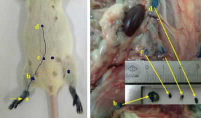
Fig. 1. The lymphatic drainage identified by blue dye in the hind foot of the mice. a, injection point; b, sentinel lymph node (popliteal); c, secondary lymph node (iliac); d, tertiary lymph node (renal).

Fig. 2. The single-photon emission computed tomography images of dynamic lymphoscintigraphy. The small particle radiotracer could be identified in the lymphoscintigraphy with the time going. R, right breast is the tumor side; L, left breast is the sound side; IMLN, internal mammary lymph node; ALN, axillary lymph node; ASLN, axillary sentinel lymph node.

Fig. 3. The combination images of single-photon emission computed tomography (SPECT)/computed tomography (CT) which helps to identify the intercostal location of internal mammary lymph nodes (IMLNs) from different anatomical aspects. ASLN, axillary sentinel lymph node.
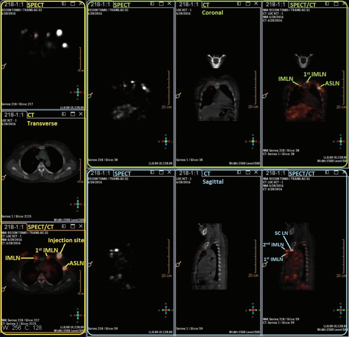
Fig. 4. The lymphoscintigraphy detected by the single-photon emission computed tomography (SPECT)/computed tomography (CT) in the next day morning about 1 hour before surgery. IMLN, internal mammary lymph node; ASLN, axillary sentinel lymph node; SCLN, supraclavicular lymph node.
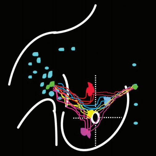
Fig. 5. The lymph in different area drain into the same internal mammary lymph node (IMLN), and then other IMLNs receive lymph from it one by one.

Fig. 6. The lymph in different area of breast drain into different internal mammary lymph nodes.
Reference
-
References
1. Connolly JL. Changes and problematic areas in interpretation of the AJCC Cancer Staging Manual, 6th Edition, for breast cancer. Arch Pathol Lab Med. 2006; 130:287–91.
Article2. Gnerlich JL, Barreto-Andrade JC, Czechura T, John JR, Turk MA, Kennedy TJ, et al. Accurate staging with internal mammary chain sentinel node biopsy for breast cancer. Ann Surg Oncol. 2014; 21:368–74.
Article3. Qiu PF, Liu YB, Wang YS. Internal mammary sentinel lymph node biopsy: abandon or persist? Onco Targets Ther. 2016; 9:3879–82.
Article4. Huang O, Wang L, Shen K, Lin H, Hu Z, Liu G, et al. Breast cancer subpopulation with high risk of internal mammary lymph nodes metastasis: analysis of 2,269 Chinese breast cancer patients treated with extended radical mastectomy. Breast Cancer Res Treat. 2008; 107:379–87.
Article5. Thorsen LB, Offersen BV, Dano H, Berg M, Jensen I, Pedersen AN, et al. DBCG-IMN: a population-based cohort study on the effect of internal mammary node irradiation in early node-positive breast cancer. J Clin Oncol. 2016; 34:314–20.
Article6. Whelan TJ, Olivotto IA, Parulekar WR, Ackerman I, Chua BH, Nabid A, et al. Regional nodal irradiation in early-stage breast cancer. N Engl J Med. 2015; 373:307–16.
Article7. Cong BB, Qiu PF, Liu YB, Zhao T, Chen P, Cao XS, et al. Validation study for the hypothesis of internal mammary sentinel lymph node lymphatic drainage in breast cancer. Oncotarget. 2016; 7:41996–2006.
Article8. Cong BB, Qiu PF, Wang YS. Internal mammary sentinel lymph node biopsy: minimally invasive staging and tailored internal mammary radiotherapy. Ann Surg Oncol. 2014; 21:2119–21.
Article9. Lyman GH, Temin S, Edge SB, Newman LA, Turner RR, Weaver DL, et al. Sentinel lymph node biopsy for patients with early-stage breast cancer: American Society of Clinical Oncology clinical practice guideline update. J Clin Oncol. 2014; 32:1365–83.
Article10. Schulze T, Mucke J, Markwardt J, Schlag PM, Bembenek A. Long-term morbidity of patients with early breast cancer after sentinel lymph node biopsy compared to axillary lymph node dissection. J Surg Oncol. 2006; 93:109–19.
Article11. Kumar A, Puri R, Gadgil PV, Jatoi I. Sentinel lymph node biopsy in primary breast cancer: window to management of the axilla. World J Surg. 2012; 36:1453–9.
Article12. Veronesi U, Marubini E, Mariani L, Valagussa P, Zucali R. The dissection of internal mammary nodes does not improve the survival of breast cancer patients. 30-year results of a randomised trial. Eur J Cancer. 1999; 35:1320–5.
Article13. Byrd DR, Dunnwald LK, Mankoff DA, Anderson BO, Moe RE, Yeung RS, et al. Internal mammary lymph node drainage patterns in patients with breast cancer documented by breast lymphoscintigraphy. Ann Surg Oncol. 2001; 8:234–40.
Article14. Qiu PF, Cong BB, Zhao RR, Yang GR, Liu YB, Chen P, et al. Internal mammary sentinel lymph node biopsy with modified injection technique: high visualization rate and accurate staging. Medicine (Baltimore). 2015; 94:e1790.15. Turner-Warwick RT. The lymphatics of the breast. Br J Surg. 1959; 46:574–82.
Article16. Cruikshank WC. The anatomy of the absorbing vessels of the human body. London: G. Nicol;1786.17. Sappey MP. Anatomie, Physiologie, Pathologie des vaisseaux Lymphatiques consideres chez L’homme at les Vertebres. Paris: A. Delahaye and E. Lecrosnier;1874.18. Pavlista D, Eliska O. Relationship between the lymphatic drainage of the breast and the upper extremity: a postmortem study. Ann Surg Oncol. 2012; 19:3410–5.
Article19. Suami H, Pan WR, Mann GB, Taylor GI. The lymphatic anatomy of the breast and its implications for sentinel lymph node biopsy: a human cadaver study. Ann Surg Oncol. 2008; 15:863–71.
Article20. Tanis PJ, Nieweg OE, Valdes Olmos RA, Kroon BB. Anatomy and physiology of lymphatic drainage of the breast from the perspective of sentinel node biopsy. J Am Coll Surg. 2001; 192:399–409.21. Yamaguchi A, Hanaoka H, Pirmettis I, Uehara T, Tsushima Y, Papadopoulos M, et al. Injection site radioactivity of (99m) Tc-labeled mannosylated dextran for sentinel lymph node mapping. Mol Pharm. 2015; 12:514–9.22. Harrell MI, Iritani BM, Ruddell A. Lymph node mapping in the mouse. J Immunol Methods. 2008; 332:170–4.
Article23. Mariani G, Erba P, Villa G, Gipponi M, Manca G, Boni G, et al. Lymphoscintigraphic and intraoperative detection of the sentinel lymph node in breast cancer patients: the nuclear medicine perspective. J Surg Oncol. 2004; 85:112–22.
Article24. Kraft O, Havel M. Sentinel lymph nodes and planar scintigraphy and SPECT/CT in various types of tumours. Estimation of some factors influencing detection success. Nucl Med Rev Cent East Eur. 2013; 16:17–25.
Article25. Brouwer OR, Vermeeren L, van der Ploeg IM, Valdes Olmos RA, Loo CE, Pereira-Bouda LM, et al. Lymphoscintigraphy and SPECT/CT in multicentric and multifocal breast cancer: does each tumour have a separate drainage pattern? Results of a Dutch multicentre study (MULTISENT). Eur J Nucl Med Mol Imaging. 2012; 39:1137–43.26. Vercellino L, Ohnona J, Groheux D, Slama A, Colletti PM, Chondrogiannis S, et al. Role of SPECT/CT in sentinel lymph node detection in patients with breast cancer. Clin Nucl Med. 2014; 39:431–6.
Article
- Full Text Links
-
- Actions
-
Cited
- CITED
-
- Close
- Share
-
- Similar articles
-
- Use of Mammary Lymphoscintigraphy and Intraoperative Radioguided Gamma Probe in Sentinel Lymph Node Biopsy of Breast Cancer
- Anatomy Versus Physiology: Is Breast Lymphatic Drainage to the Internal Thoracic (Internal Mammary) Lymphatic System Clinically Relevant?
- Camparison of the Efficiency for Tc-99m Tin-colloid and Tc-99m Phytate in Sentinel Node Detection in Breast Cancer Patients
- A Case of Anaphylactic Reactions to Isosulfan Blue Dye in A Cervical Cancer Patient
- Tc-99m Diphosphonate as a Potential Radiotracer to Detect Sentinel Lymph Nodes in Patients with Breast Cancer
