Diagnosis of tooth wear
- Affiliations
-
- 1Department of Prosthodontics and Research Institute of Oral Science, College of Dentistry, Gangneung-Wonju National University, Gangneung, Republic of Korea. lila@gwnu.ac.kr
- KMID: 2463620
- DOI: http://doi.org/10.14368/jdras.2019.35.3.113
Abstract
- Tooth wear is gradually increasing with increasing life expectancy. In particular, it is important to establish a treatment plan in the early stages so that it does not proceed to moderate or severe wear stages. It is essential to diagnose tooth wear accurately in order to plan a treatment for it. There are many risk factors including age, diet, and drugs which affects tooth wear. For the diagnosis of a tooth wear, appropriate index and evaluation method should be used. There were various tooth wear indices such as TWI, Lussi index, BEWE, and TWES. The evaluation method includes clinical examination, dental cast examination and clinical photographs. Recently, a 3D scanner is being used to assess tooth wear. The risk factors, tooth wear evaluation system, the methods of measuring tooth wear, and related literature were reviewed. The strengths and weaknesses of each index and evaluation methods were compared to derive a proper way to diagnose tooth wear.
Keyword
Figure
-
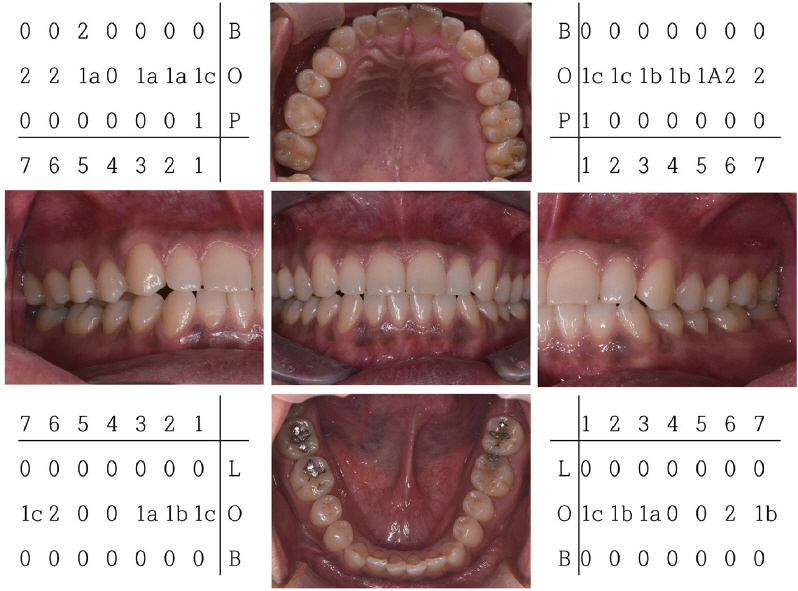
Fig. 1 Example of tooth wear scoring with clinical photographs by TWES (tooth wear evaluation system) (B: buccal surface, O: occlusal surface, P:palatal surface, L: lingual surface). All TWES scores were 1, 2, which represented the early stage of generalized occlusal wear. According to the TWES guidelines, counselling/monitoring is recommended.

Fig. 2 Example of tooth wear scoring with dental cast by TWES (tooth wear evaluation system) (B: buccal surface, O: occlusal surface, P: palatal surface, L: lingual surface). TWES scores in the cast are generally increased compared to that of the clinical examination. It appears to have increased because the surface texture can be observed well in the cast.
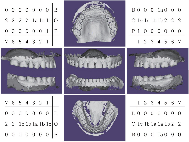
Fig. 3 Example of tooth wear scoring with 3D scanning of dental cast by TWES (tooth wear evaluation system) (B: buccal surface, O: occlusal surface, P: palatal surface, L: lingual surface). TWES scores are increased compared to the scores measured in the cast. This is assumed to be due to differences in resolution and scan errors. Note the indeterminate cervical abrasion in maxillary right second premolar. Because the scan was scanned, the errors in the scan were replicated in scanned data.
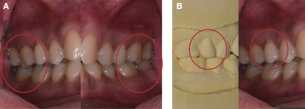
Fig. 4 Possible errors. (A) Maxillary second molars are covered by soft tissue, (B) Void in buccal surface of maxillary right second premolar
Reference
-
References
1. Johansson AK, Omar R, Carlsson GE, Johansson A. Dental erosion and its growing importance in clinical practice:from past to present. Int J Dent. 2012; 2012:632907. DOI: 10.1155/2012/632907. PMID: 22505907. PMCID: PMC3312266.2. Smith BG, Knight JK. An index for measuring the wear of teeth. Br Dent J. 1984; 156:435–8. DOI: 10.1038/sj.bdj.4805394. PMID: 6590081.3. Wetselaar P, Faris A, Lobbezoo F. A plea for the development of an universally accepted modular tooth wear evaluation system. BMC Oral Health. 2016; 16:115. DOI: 10.1186/s12903-016-0309-6. PMID: 27809895. PMCID: PMC5094144.4. Hattab FN, Yassin OM. Etiology and diagnosis of tooth wear:a literature review and presentation of selected cases. Int J Prosthodont. 2000; 13:101–7. PMID: 11203616.5. Lussi A, Carvalho TS. Erosive tooth wear:a multifactorial condition of growing concern and increasing knowledge. Monogr Oral Sci. 2014; 25:1–15. DOI: 10.1159/000360380. PMID: 24993253.6. Smith BG, Bartlett DW, Robb ND. The prevalence, etiology and management of tooth wear in the United Kingdom. J Prosthet Dent. 1997; 78:367–72. DOI: 10.1016/S0022-3913(97)70043-X. PMID: 9338867.7. Wetselaar P, Vermaire JH, Visscher CM, Lobbezoo F, Schuller AA. The Prevalence of Tooth Wear in the Dutch Adult Population. Caries Res. 2016; 50:543–50. DOI: 10.1159/000447020. PMID: 27694757. PMCID: PMC5296789.8. Kitasako Y, Sasaki Y, Takagaki T, Sadr A, Tagami J. Age-specific prevalence of erosive tooth wear by acidic diet and gastroesophageal reflux in Japan. J Dent. 2015; 43:418–23. DOI: 10.1016/j.jdent.2015.02.004. PMID: 25684603.9. Wei Z, Du Y, Zhang J, Tai B, Du M, Jiang H. Prevalence and Indicators of Tooth Wear among Chinese Adults. PLoS One. 2016; 11:e0162181. DOI: 10.1371/journal.pone.0162181. PMID: 27583435. PMCID: PMC5008775.10. Holbrook WP, Arnadottir IB, Kay EJ. Prevention. Part 3:prevention of tooth wear. Br Dent J. 2003; 195:75–81. DOI: 10.1038/sj.bdj.4810331. PMID: 12881743.11. Hugoson A, Bergendal T, Ekfeldt A, Helkimo M. Prevalence and severity of incisal and occlusal tooth wear in an adult Swedish population. Acta Odontol Scand. 1988; 46:255–65. DOI: 10.3109/00016358809004775. PMID: 3264985.12. Van't Spijker A, Rodriguez JM, Kreulen CM, Bronkhorst EM, Bartlett DW, Creugers NH. Prevalence of tooth wear in adults. Int J Prosthodont. 2009; 22:35–42. PMID: 19260425.13. Vered Y, Lussi A, Zini A, Gleitman J, Sgan-Cohen HD. Dental erosive wear assessment among adolescents and adults utilizing the basic erosive wear examination (BEWE) scoring system. Clin Oral Investig. 2014; 18:1985–90. DOI: 10.1007/s00784-013-1175-0. PMID: 24420504.14. Moazzez R, Bartlett D, Anggiansah A. Dental erosion, gastro-oesophageal reflux disease and saliva:how are they related? J Dent. 2004; 32:489–94. DOI: 10.1016/j.jdent.2004.03.004. PMID: 15240067.15. Al-Omiri MK, Sghaireen MG, Alzarea BK, Lynch E. Quantification of incisal tooth wear in upper anterior teeth:conventional vs new method using toolmakers microscope and a three-dimensional measuring technique. J Dent. 2013; 41:1214–21. DOI: 10.1016/j.jdent.2013.08.022. PMID: 24012517.16. Tantbirojn D, Pintado MR, Versluis A, Dunn C, Delong R. Quantitative analysis of tooth surface loss associated with gastroesophageal reflux disease:a longitudinal clinical study. J Am Dent Assoc. 2012; 143:278–85. DOI: 10.14219/jada.archive.2012.0153. PMID: 22383209.17. Smith BG, Knight JK. A comparison of patterns of tooth wear with aetiological factors. Br Dent J. 1984; 157:16–9. DOI: 10.1038/sj.bdj.4805401. PMID: 6588978.18. Bartlett DW, Lussi A, West NX, Bouchard P, Sanz M, Bourgeois D. Prevalence of tooth wear on buccal and lingual surfaces and possible risk factors in young European adults. J Dent. 2013; 41:1007–13. DOI: 10.1016/j.jdent.2013.08.018. PMID: 24004965.19. Nolen-Hoeksema S, Wisco BE, Lyubomirsky S. Rethinking Rumination. Perspect Psychol Sci. 2008; 3:400–24. DOI: 10.1111/j.1745-6924.2008.00088.x. PMID: 26158958.20. Wiegand A, Attin T. Occupational dental erosion from exposure to acids:a review. Occup Med (Lond). 2007; 57:169–76. DOI: 10.1093/occmed/kql163. PMID: 17307767.21. Nunn J, Shaw L, Smith A. Tooth wear-dental erosion. Br Dent J. 1996; 180:349–52. DOI: 10.1038/sj.bdj.4809084. PMID: 8664093.22. Craddock HL, Youngson CC, Manogue M, Blance A. Occlusal changes following posterior tooth loss in adults. Part 2. Clinical parameters associated with movement of teeth adjacent to the site of posterior tooth loss. J Prosthodont. 2007; 16:495–501. DOI: 10.1111/j.1532-849X.2007.00223.x. PMID: 17672834.23. Lussi A, Schaffner M, Hotz P, Suter P. Dental erosion in a population of Swiss adults. Community Dent Oral Epidemiol. 1991; 19:286–90. DOI: 10.1111/j.1600-0528.1991.tb00169.x. PMID: 1742995.24. Bartlett D, Ganss C, Lussi A. Basic Erosive Wear Examination (BEWE):a new scoring system for scientific and clinical needs. Clin Oral Investig. 2008; 12(Suppl 1):S65–8. DOI: 10.1007/s00784-007-0181-5. PMID: 18228057. PMCID: PMC2238785.25. Wetselaar P, Lobbezoo F. The tooth wear evaluation system:a modular clinical guideline for the diagnosis and management planning of worn dentitions. J Oral Rehabil. 2016; 43:69–80. DOI: 10.1111/joor.12340. PMID: 26333037.26. Bartlett DW. Retrospective long term monitoring of tooth wear using study models. Br Dent J. 2003; 194:211–3. discussion 204. DOI: 10.1038/sj.bdj.4809914. PMID: 12627200.27. Vervoorn-Vis GM, Wetselaar P, Koutris M, Visscher CM, Evalahti M, Ahlberg J, Lobbezoo F. Assessment of the progression of tooth wear on dental casts. J Oral Rehabil. 2015; 42:600–4. DOI: 10.1111/joor.12292. PMID: 25752246.28. Alaraudanjoki V, Saarela H, Pesonen R, Laitala ML, Kiviahde H, Tjaderhane L, Lussi A, Pesonen P, Anttonen V. Is a Basic Erosive Wear Examination (BEWE) reliable for recording erosive tooth wear on 3D models? J Dent. 2017; 59:26–32. DOI: 10.1016/j.jdent.2017.02.001. PMID: 28189719.29. Hove LH, Mulic A, Tveit AB, Stenhagen KR, Skaare AB, Espelid I. Registration of dental erosive wear on study models and intra-oral photographs. Eur Arch Paediatr Dent. 2013; 14:29–34. DOI: 10.1007/s40368-012-0004-5. PMID: 23532811.30. Holbrook WP, Ganss C. Is diagnosing exposed dentine a suitable tool for grading erosive loss? Clin Oral Investig. 2008; 12(Suppl 1):S33–9. DOI: 10.1007/s00784-007-0174-4. PMID: 18228063. PMCID: PMC2238781.31. Wetselaar P, Lobbezoo F, Koutris M, Visscher CM, Naeije M. Reliability of an occlusal and nonocclusal tooth wear grading system:clinical use versus dental cast assessment. Int J Prosthodont. 2009; 22:388–90. PMID: 19639077.32. Ganss C, Klimek J, Giese K. Dental erosion in children and adolescents-a cross-sectional and longitudinal investigation using study models. Community Dent Oral Epidemiol. 2001; 29:264–71. DOI: 10.1034/j.1600-0528.2001.290405.x. PMID: 11515640.33. Wetselaar P, Wetselaar-Glas MJ, Koutris M, Visscher CM, Lobbezoo F. Assessment of the amount of tooth wear on dental casts and intra-oral photographs. J Oral Rehabil. 2016; 43:615–20. DOI: 10.1111/joor.12405. PMID: 27132187.34. Marro F, De Lat L, Martens L, Jacquet W, Bottenberg P. Monitoring the progression of erosive tooth wear (ETW) using BEWE index in casts and their 3D images:A retrospective longitudinal study. J Dent. 2018; 73:70–5. DOI: 10.1016/j.jdent.2018.04.008. PMID: 29660487.35. Ahmed KE, Whitters J, Ju X, Pierce SG, MacLeod CN, Murray CA. Clinical Monitoring of Tooth Wear Progression in Patients over a Period of One Year Using CAD/CAM. Int J Prosthodont. 2017; 30:153–5. DOI: 10.11607/ijp.4990. PMID: 28267825.36. Peters MC, Delong R, Pintado MR, Pallesen U, Qvist V, Douglas WH. Comparison of two measurement techniques for clinical wear. J Dent. 1999; 27:479–85. DOI: 10.1016/S0300-5712(99)00027-5. PMID: 10507203.37. Carvalho TS, Colon P, Ganss C, Huysmans MC, Lussi A, Schlueter N, Schmalz G, Shellis RP, Tveit AB, Wiegand A. Consensus report of the European Federation of Conservative Dentistry:erosive tooth wear-diagnosis and management. Clin Oral Investig. 2015; 19:1557–61. DOI: 10.1007/s00784-015-1511-7. PMID: 26121968.
- Full Text Links
-
- Actions
-
Cited
- CITED
-
- Close
- Share
-
- Similar articles
-
- The interpretation of tooth wear - literature review
- Review of Previous Studies on Dental Wear Method
- A novel 3D scan method to quantify teeth wear
- Oral rehabilitation of excessive tooth wear patient usingzirconia fixed prosthesis with increased vertical dimension
- Full mouth rehabilitation of the patient with severe tooth loss and tooth wear with vertical dimension gaining: A case report
