J Adv Prosthodont.
2019 Oct;11(5):239-246. 10.4047/jap.2019.11.5.239.
Proposal for a gingival shade guide based on in vivo spectrophotometric measurements
- Affiliations
-
- 1Department of Dentistry, School of Medicine, Campus Miguel de Unamuno, University of Salamanca, Spain. crisgodent@hotmail.com
- KMID: 2462106
- DOI: http://doi.org/10.4047/jap.2019.11.5.239
Abstract
- PURPOSE
The purpose of this study was to propose and assess a shade guide for pink gingival aesthetics using a Spanish population sample.
MATERIALS AND METHODS
The L*, C*, h, a* and b* coordinates of 259 participants were measured using a spectrophotometer in 3 standardized points along the attached gingiva of the maxillary central incisors. A hierarchical clustering analysis was applied to obtain separate solutions regarding the number of shade tabs. For each of the solutions obtained, color differences (ΔE*) were calculated using the CIELab and CIEDE2000 formulas, and the proposed shade guide was selected considering (1) the color differences between tabs and (2) the coverage error of each of the solutions.
RESULTS
The proposed shade guide consisted of 8 gingival shade tabs and achieved CIELab and CIEDE2000 coverage errors of less than the respective 50:50% acceptability thresholds (ΔE*=4.6 units and ΔE₀₀=4.1). The coordinates for the various gingival shade tabs were as follows: Tab 1: L*43.3, a*21.9, b*12.3 (1.6); Tab 2: L*42.9, a*34.1, b*19.1; Tab 3: L*46.5, a*25.8, b*10.9; Tab 4: L*46.5, a*27.3, b*15.1; Tab 5: L*49.6, a*23.5, b*16.8; Tab 6: L*51.5, a*19.7, b*13.6; Tab 7: L*55.9, a*22.0, b* 15.0; and Tab 8: L*56.0, a*19.9, b*18.8.
CONCLUSION
The CIELab and CIEDE2000 coverage errors for the 8 shade tabs of the proposed gingival shade guide were significantly lower than those of other guides. Therefore, despite the limitations of this study, the proposed guide is more appropriate for matching gingival shade in the Spanish general population.
Figure
-
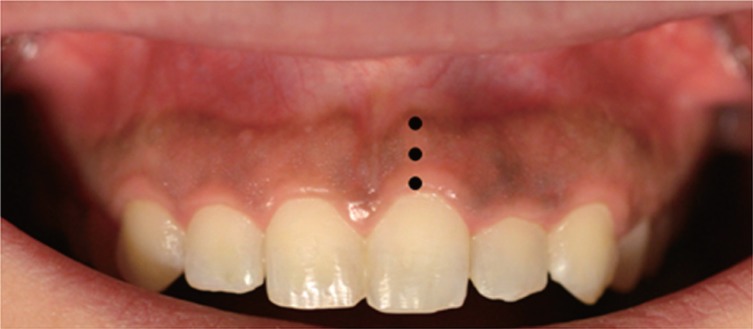
Fig. 1 Measuring points distributed along the keratinized gingiva.

Fig. 2 CIELab and CIEDE2000 coverage error values for the different solutions.
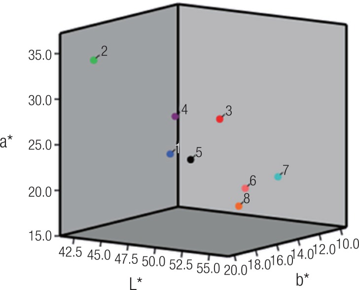
Fig. 3 Scatter plot of the mean values of L*, a*, and b* for the 8 cluster group centroids.
Reference
-
1. Miller PD Jr. A classification of marginal tissue recession. Int J Periodontics Restorative Dent. 1985; 5:8–13.2. Miller PD Jr. Root coverage grafting for regeneration and aesthetics. Periodontol 2000. 1993; 1:118–127.
Article3. Ghinea R, Herrera LJ, Pérez MM, Ionescu AM, Paravina RD. Gingival shade guides: Colorimetric and spectral modeling. J Esthet Restor Dent. 2018; 30:E31–E38. PMID: 29667787.
Article4. Gómez-Polo C, Montero J, Gómez-Polo M, Martín Casado AM. Clinical study on natural gingival color. Odontology. 2019; 107:80–89. PMID: 29845402.
Article5. Sala L, Carrillo-de-Albornoz A, Martín C, Bascones-Martínez A. Factors involved in the spectrophotometric measurement of soft tissue: A clinical study of interrater and intrarater reliability. J Prosthet Dent. 2015; 113:558–564. PMID: 25794908.
Article6. Gómez-Polo C, Gómez-Polo M, Martínez Vázquez de Parga JA, Celemín-Viñuela A. Clinical study of the 3D-master color system among the Spanish population. J Prosthodont. 2018; 27:708–715. PMID: 28084028.
Article7. Yuan JC, Brewer JD, Monaco EA Jr, Davis EL. Defining a natural tooth color space based on a 3-dimensional shade system. J Prosthet Dent. 2007; 98:110–119. PMID: 17692592.
Article8. Ahn JS, Lee YK. Color distribution of a shade guide in the value, chroma, and hue scale. J Prosthet Dent. 2008; 100:18–28. PMID: 18589070.
Article9. Khurana PR, Thomas PV, Rao SV, Balamuragan R, Singh MP. A clinical study to correlate maxillary anterior natural teeth with that of the commercially available acrylic and porcelain shade guides. J Contemp Dent Pract. 2013; 14:427–433. PMID: 24171984.
Article10. Gómez-Polo C, Montero J, Gómez-Polo M, de Parga JA, Celemin-Viñuela A. Natural Tooth color estimation based on age and gender. J Prosthodont. 2017; 26:107–114. PMID: 26371458.
Article11. Gozalo-Diaz D, Johnston WM, Wee AG. Estimating the color of maxillary central incisors based on age and gender. J Prosthet Dent. 2008; 100:93–98. PMID: 18672125.
Article12. Eiffler C, Cevirgen E, Helling S, Zornek J, Pritsch M, Hassel AJ. Differences in lightness, chroma, and hue in the anterior teeth of quinquagenarians and septuagenarians. Clin Oral Investig. 2010; 14:587–591.
Article13. Bayindir F, Bayindir YZ, Gozalo-Diaz DJ, Wee AG. Coverage error of gingival shade guide systems in measuring color of attached anterior gingiva. J Prosthet Dent. 2009; 101:46–53. PMID: 19105991.
Article14. Huang JW, Chen WC, Huang TK, Fu PS, Lai PL, Tsai CF, Hung CC. Using a spectrophotometric study of human gingival colour distribution to develop a shade guide. J Dent. 2011; 39:e11–e16. PMID: 22005337.
Article15. Ito M, Marx DB, Cheng AC, Wee AG. Proposed shade guide for attached gingiva-a pilot study. J Prosthodont. 2015; 24:182–187. PMID: 25081432.
Article16. Sproull RC. Color matching in dentistry: Part I. The three-dimensional nature of color. J Prosthet Dent. 1973; 29:416–424. PMID: 4511779.
Article17. CIE (Commission Internationale de l'Eclairage). Annuaire, Roster, Register, Annexe au Bulletin CIE. Parıs: Bureau Central de la CIE;1976. (PROCLUS, patrimonio Conicet).18. Gómez-Polo C, Montero J, Gómez-Polo M, Martin Casado A. Comparison of the CIELab and CIEDE 2000 color difference formulas on gingival color space. J Prosthodont. 2017; 12. 22. DOI: 10.1111/jopr.12717. [Epub ahead of print].19. Amer RS, Chandrasekaran I, Johnston WM. Illuminant effect on the coverage error of a gingiva-colored composite resin shade guide. J Prosthet Dent. 2016; 116:770–776. PMID: 27312656.
Article20. Pérez MM, Ghinea R, Herrera LJ, Carrillo F, Ionescu AM, Paravina RD. Color difference thresholds for computer-simulated human Gingiva. J Esthet Restor Dent. 2018; 30:E24–E30. PMID: 29476587.
Article21. Lee YK. Comparison of CIELAB DeltaE(*) and CIEDE2000 color-differences after polymerization and thermocycling of resin composites. Dent Mater. 2005; 21:678–682. PMID: 15978278.22. Ren J, Lin H, Huang Q, Zheng G. Determining color difference thresholds in denture base acrylic resin. J Prosthet Dent. 2015; 114:702–708. PMID: 26277020.
Article23. Luo MR, Cui G, Rigg B. The development of the CIE 2000 colour-difference formula: CIEDE2000. Color Res Appl. 2001; 26:340–350.
Article24. CIE Publication 142-2001. ISO/CIE 11664-6: 2014 (CIE S 014-6/E:2013).25. CIE 15. Technical Report: Colorimetry. 3rd ed. 2004.26. Sharma G, Wu W, Dalal EN. The CIEDE2000 color-difference formula: Implementation notes, supplementary test data, and mathematical observations. Color Res Appl. 2005; 30:21–30.
Article27. Pecho OE, Pérez MM, Ghinea R, Della Bona A. Lightness, chroma and hue differences on visual shade matching. Dent Mater. 2016; 32:1362–1373. PMID: 27614615.
Article28. Sharma G. The CIEDE2000 Color-difference formula: Implementation notes, supplementary test data, and mathematical observations. Excel spreadsheet implementation of the CIEDE2000 color-difference formula (including test data). Available at: http://www2.ece.rochester.edu/~gsharma/ciede2000/.29. Ishikawa N. Study on measuring method of gingival color. Bull Tokyo Med Dent Univ. 1961; 8:115.30. Baumgartner WJ, Weis RP, Reyher JL. The diagnostic value of redness in gingivitis. J Periodontol. 1966; 37:294–297. PMID: 5220896.
Article31. Dummett CO, Sakumura JS, Barens G. The relationship of facial skin complexion to oral mucosa pigmentation and tooth color. J Prosthet Dent. 1980; 43:392–396. PMID: 6928477.
Article32. Kleinheinz J, Büchter A, Fillies T, Joos U. Vascular basis of mucosal color. Head Face Med. 2005; 1:4. PMID: 16270929.
Article33. Heydecke G, Schnitzer S, Türp JC. The color of human gingiva and mucosa: visual measurement and description of distribution. Clin Oral Investig. 2005; 9:257–265.
Article34. Loe H, Silness J. Periodontal disease in pregnancy. I. Prevalence and severity. Acta Odontol Scand. 1963; 21:533–551. PMID: 14121956.35. Lang NP, Tonetti MS. Periodontal risk assessment (PRA) for patients in supportive periodontal therapy (SPT). Oral Health Prev Dent. 2003; 1:7–16. PMID: 15643744.36. Jung RE, Sailer I, Hämmerle CH, Attin T, Schmidlin P. In vitro color changes of soft tissues caused by restorative materials. Int J Periodontics Restorative Dent. 2007; 27:251–257. PMID: 17694948.37. Bressan E, Paniz G, Lops D, Corazza B, Romeo E, Favero G. Influence of abutment material on the gingival color of implant-supported all-ceramic restorations: a prospective multicenter study. Clin Oral Implants Res. 2011; 22:631–637. PMID: 21070378.
Article38. Happe A, Schulte-Mattler V, Strassert C, Naumann M, Stimmelmayr M, Zoller JE, Rothamel D. In vitro color changes of soft tissues caused by dyed fluorescent zirconia and nondyed, nonfluorescent zirconia in thin mucosa. Int J Periodontics Restorative Dent. 2013; 33:e1–e8. PMID: 23342353.
Article39. Sailer I, Fehmer V, Ioannidis A, Hämmerle CH, Thoma DS. Threshold value for the perception of color changes of human gingiva. Int J Periodontics Restorative Dent. 2014; 34:757–762. PMID: 25411730.
Article40. Brewer JD, Wee A, Seghi R. Advances in color matching. Dent Clin North Am. 2004; 48:v341–358.
Article41. Hugo B, Witzel T, Klaiber B. Comparison of in vivo visual and computer-aided tooth shade determination. Clin Oral Investig. 2005; 9:244–250.
Article42. Gozalo-Diaz DJ, Lindsey DT, Johnston WM, Wee AG. Measurement of color for craniofacial structures using a 45/0-degree optical configuration. J Prosthet Dent. 2007; 97:45–53. PMID: 17280891.
Article43. Everitt BS, Landau S, Leese M, Stahl D. Cluster Analysis. 5th ed. Wiley;2011.44. Analoui M, Papkosta E, Cochran M, Matis B. Designing visually optimal shade guides. J Prosthet Dent. 2004; 92:371–376. PMID: 15507911.
Article45. Ward JH Jr. Hierarchical grouping to optimize an objective function. J American Statistical Assoc. 1963; 58:236–244.
Article46. Thoma DS, Ioannidis A, Fehmer V, Michelotti G, Jung RE, Sailer I. Threshold values for the perception of color changes in human teeth. Int J Periodontics Restorative Dent. 2016; 36:777–783. PMID: 27922642.
Article47. Sarmast ND, Angelov N, Ghinea R, Powers JM, Paravina RD. Color compatibility of gingival shade guides and gingiva-colored dental materials with healthy human gingiva. Int J Periodontics Restorative Dent. 2018; 38:397–403. PMID: 29641629.
Article48. Trushkowsky RD. How a spectrophotometer can help you achieve esthetic shade matching. Compend Contin Educ Dent. 2003; 24:60–66. PMID: 12619260.49. Joiner A. Tooth colour: a review of the literature. J Dent. 2004; 32:3–12.
Article50. Ho DK, Ghinea R, Herrera LJ, Angelov N, Paravina RD. Color range and color distribution of healthy human gingiva: a prospective clinical study. Sci Rep. 2015; 5:18498. PMID: 26691598.
Article51. Barghi N, Pedrero JA, Bosch RR. Effects of batch variation on shade of dental porcelain. J Prosthet Dent. 1985; 54:625–627. PMID: 3903130.
- Full Text Links
-
- Actions
-
Cited
- CITED
-
- Close
- Share
-
- Similar articles
-
- Spectrophotometric evaluation of restorative composite shades and their match with a classical shade guide
- Shade comparative analysis of natural tooth measured by visual and spectrophotometric methods
- ANALYSIS OF OPTICAL TRANSMISSION CHARATERISTICS BY VISIBLE LIGHT INTO SHADE GUIDES
- Influence of the shade guide design on color matching
- The reproducibility of various porcelain color selection systems using spectrophotometry
