Ann Dermatol.
2019 Dec;31(6):666-668. 10.5021/ad.2019.31.6.666.
Localized Cutaneous Argyria Mimicking Blue Nevus after Wearing Earrings
- Affiliations
-
- 1Department of Dermatology, Samsung Changwon Hospital, Sungkyunkwan University School of Medicine, Changwon, Korea. rkw3840@gmail.com
- KMID: 2461990
- DOI: http://doi.org/10.5021/ad.2019.31.6.666
Abstract
- Localized cutaneous argyria is a rare condition in which the skin changes into blue-grey spots due to the absorption of silver. The lesions need to be differentiated from other pigmentary disorders and require radiographic and histological examination for more accurate diagnosis. Scanning electron microscopy and energy dispersive x-ray spectroscopy can be a confirmatory tool in the evaluation of silver elements in biopsy tissue. This report shows the localized cutaneous argyria in earlobe of a 21-year-old woman who wears silver earrings for 10 years.
MeSH Terms
Figure
-
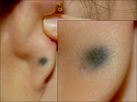
Fig. 1 The patient presented solitary bluish macule on right posterior earlobe.

Fig. 2 Microscopic examination of a biopsy specimen from a right earlobe. (A) Discrete, fine, dark granules and deposition of foreign substance on the basement membrane of eccrine sweat glands (H&E, ×400). (B) Deposition of brown fiber-like foreign substance (arrowheads) on the dermal elastic and collagen fibers (H&E, ×400). (C) Scanning electron microscopy demonstrates small white particles (arrow). BSE: backscattered electrons mode. (D) Energy dispersive x-ray spectroscopy analysis identified the particles as minute fragments of silver (Ag) and confirms admixed silica (Si).
Reference
-
1. Morton CA, Fallowfield M, Kemmett D. Localized argyria caused by silver earrings. Br J Dermatol. 1996; 135:484–485.
Article2. McClain CM, Kantrow SM, Abraham JL, Price J, Parker ER, Robbins JB. Localized cutaneous argyria: two case reports and clinicopathologic review. Am J Dermatopathol. 2013; 35:e115–e118.3. Kwon IH, Ryu TH, Kye H, Kim DH, Choi JE, Seo SH, et al. Cutaneous argyria confirmed by scanning electron microscopy and energy dispersive x-ray (SEM-EDX) spectroscopy. Korean J Dermatol. 2014; 66:Suppl 1. 506–507.4. Park MY, Jin H, You HS, Shim WH, Kim JM, Kim GW, et al. Localized argyria Troublesome side effect of acupuncture. Korean J Dermatol. 2016; 68:Suppl 1. 304–305.5. Garcias-Ladaria J, Hernandez-Bel P, Torregrosa-Calatayud JL, Martínez-Aparicio A. Localized cutaneous argyria: a report of 2 cases. Actas Dermosifiliogr. 2013; 104:253–254.
Article6. Hristov AC, High WA, Golitz LE. Localized cutaneous argyria. J Am Acad Dermatol. 2011; 65:660–661.
Article7. Park SW, Kim JH, Shin HT, Lee KT, Lee JH, Lee DY, et al. An effective modality for argyria treatment: Q-switched 1,064-nm Nd:YAG laser. Ann Dermatol. 2013; 25:511–512.
Article
