Lateral Ventricular Meningioma Presenting with Intraventricular Hemorrhage
- Affiliations
-
- 1Department of Neurosurgery, Pusan National University Hospital, Pusan National University School of Medicine, Busan, Korea. neurocha@hanmail.net
- KMID: 2461190
- DOI: http://doi.org/10.14791/btrt.2019.7.e41
Abstract
- A 35-year-old female visited emergency department for a sudden onset of headache with vomiting after management for abortion at local department. Neurological examination revealed drowsy mentality without focal neurological deficits. CT showed 3.2×3.4 cm hyperdense intraventricular mass with intraventricular hemorrhage. The intraventricular hemorrhage was found in lateral, 3rd, and 4th ventricles. MRI showed well enhancing intraventricular mass abutting choroid plexus in the trigone of the right lateral ventricle. CT angiography showed tortuous prominent arteries from choroidal artery in tumor. Her neurological status deteriorated to stupor and contralateral hemiparesis during planned preoperative workup. Urgent transtemporal and transcortical approach with decompressive craniectomy for removal of intraventricular meningioma with hemorrhage was done. Grossly total removal of ventricular mass was achieved. Pathological finding was meningotheliomatous meningioma of World Health Organization (WHO) grade I. The patient recovered to alert mentality and no motor deficit after intensive care for increased intracranial pressure. However, visual field defect was developed due to posterior cerebral artery territory infarction. The visual deficit did not resolve during follow up period. Lateral ventricular meningioma with spontaneous intraventricular hemorrhage in pregnant woman is very uncommon. We report a surgical case of lateral ventricular meningioma with rapid neurological deterioration for intraventricular hemorrhage.
Keyword
MeSH Terms
-
Adult
Angiography
Arteries
Choroid
Choroid Plexus
Critical Care
Decompressive Craniectomy
Emergency Service, Hospital
Female
Follow-Up Studies
Fourth Ventricle
Headache
Hemorrhage*
Humans
Infarction
Intracranial Pressure
Lateral Ventricles
Magnetic Resonance Imaging
Meningioma*
Neurologic Examination
Paresis
Posterior Cerebral Artery
Pregnancy
Pregnant Women
Stupor
Visual Fields
Vomiting
World Health Organization
Figure
-
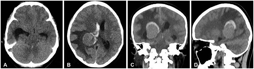
Fig. 1 Preoperative axial (A, B), coronal (C), and sagittal (D) CT showing a homogenous mass in trigone of right lateral ventricle surrounded by hemorrhage with enlargement of ipsilateral temporal horn.
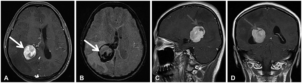
Fig. 2 Preoperative axial (A), susceptibility weighted imaging (B), sagittal (C), and coronal (D) enhanced MRI showing homogenous enhancing mass in right trigone with hemorrhage (arrow) from the tumor periphery.
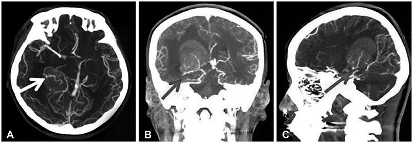
Fig. 3 Axial (A), coronal (B), and sagittal (C) enhanced CT angiography showing enlarged tortuous feeding artery (arrow) from anterior choroidal artery.
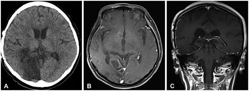
Fig. 4 Postoperative CT (A) and MRI (B, C) showing total removal of trigonal meningioma with infarction of right occipital lobe for occlusion of posterior cerebral artery from preoperative transient transtentorial herniation.
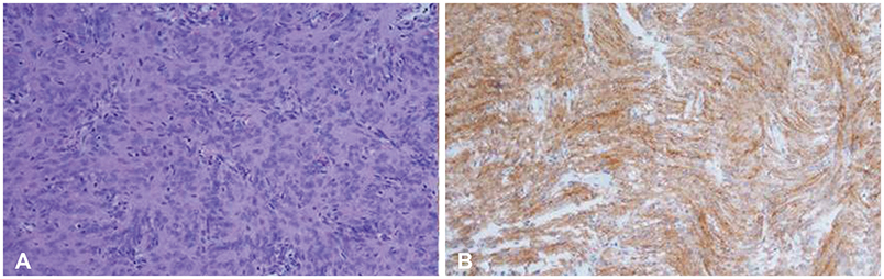
Fig. 5 Histopathological findings. World Health Organization grade I meningotheliomatous meningioma with meningothelial cells exhibiting whorled growth (hematoxylin and eosin staining, ×200) (A) and epithelial membrane antigen (+) (×200) (B).
Reference
-
1. Nakamura M, Roser F, Bundschuh O, Vorkapic P, Samii M. Intraventricular meningiomas: a review of 16 cases with reference to the literature. Surg Neurol. 2003; 59:491–503. discussion 503-4.
Article2. Kim EY, Kim ST, Kim HJ, Jeon P, Kim KH, Byun HS. Intraventricular meningiomas: radiological findings and clinical features in 12 patients. Clin Imaging. 2009; 33:175–180.
Article3. Lyngdoh BT, Giri PJ, Behari S, Banerji D, Chhabra DK, Jain VK. Intraventricular meningiomas: a surgical challenge. J Clin Neurosci. 2007; 14:442–448.
Article4. Bertalanffy A, Roessler K, Koperek O, et al. Intraventricular meningiomas: a report of 16 cases. Neurosurg Rev. 2006; 29:30–35.
Article5. Ma J, Cheng L, Wang G, Lin S. Surgical management of meningioma of the trigone area of the lateral ventricle. World Neurosurg. 2014; 82:757–769.
Article6. Bosnjak R, Derham C, Popović M, Ravnik J. Spontaneous intracranial meningioma bleeding: clinicopathological features and outcome. J Neurosurg. 2005; 103:473–484.
Article7. Niiro M, Ishimaru K, Hirano H, Yunoue S, Kuratsu J. Clinico-pathological study of meningiomas with haemorrhagic onset. Acta Neurochir (Wien). 2003; 145:767–772.
Article8. Vij M, Jaiswal S, Jaiswal AK, Kumar S, Behari S. Meningioma with hemorrhagic onset: two case reports. J Cancer Res Ther. 2012; 8:145–147.
Article9. Kim JH, Gwak HS, Hong EK, Bang CW, Lee SH, Yoo H. A case of benign meningioma presented with subdural hemorrhage. Brain Tumor Res Treat. 2015; 3:30–33.
Article10. Helle TL, Conley FK. Haemorrhage associated with meningioma: a case report and review of the literature. J Neurol Neurosurg Psychiatry. 1980; 43:725–729.
Article11. Gruszkiewicz J, Doron Y, Gellei B, Peyser E. Massive intracerebral bleeding due to supratentorial meningioma. Neurochirurgia (Stuttg). 1969; 12:107–111.
Article12. Bloomgarden GM, Byrne TN, Spencer DD, Heafner MD. Meningioma associated with aneurysm and subarachnoid hemorrhage: case report and review of the literature. Neurosurgery. 1987; 20:24–26.
Article13. Martínez-Lage JF, Poza M, Martínez M, Esteban JA, Antúnez MC, Sola J. Meningiomas with haemorrhagic onset. Acta Neurochir (Wien). 1991; 110:129–132.
Article14. Kim DG, Park CK, Paek SH, et al. Meningioma manifesting intracerebral haemorrhage: a possible mechanism of haemorrhage. Acta Neurochir (Wien). 2000; 142:165–168.
Article15. Lusis EA, Scheithauer BW, Yachnis AT, et al. Meningiomas in pregnancy: a clinicopathologic study of 17 cases. Neurosurgery. 2012; 71:951–961.16. Krause DN, Duckles SP, Pelligrino DA. Influence of sex steroid hormones on cerebrovascular function. J Appl Physiol (1985). 2006; 101:1252–1261.
Article
- Full Text Links
-
- Actions
-
Cited
- CITED
-
- Close
- Share
-
- Similar articles
-
- Intraventricular Hemorrhage Caused by Lateral Ventricular Meningioma: A Case Report
- A Case of Intraventricular Meningioma Acompanied by Intraventricular Hematoma and Subarachnoid Hemorrhage: Case Report
- A Case of Giant Intraventricular Meningioma
- Change of the Ventricular System Following Intraventricular Injection of the Blood
- Two Cases of Intraventricular Meningiomas
