Korean Circ J.
2019 Nov;49(11):1112-1113. 10.4070/kcj.2019.0244.
Images of Mitral Valve Perforation due to Atrial Septal Occluder Device
- Affiliations
-
- 1Department of Thoracic and Cardiovascular Surgery, Sejong General Hospital, Bucheon, Korea.
- 2Division of Cardiovascular Disease, Sejong General Hospital, Bucheon, Korea. learnbyliving9@gmail.com
- KMID: 2460534
- DOI: http://doi.org/10.4070/kcj.2019.0244
Abstract
- No abstract available.
MeSH Terms
Figure
-
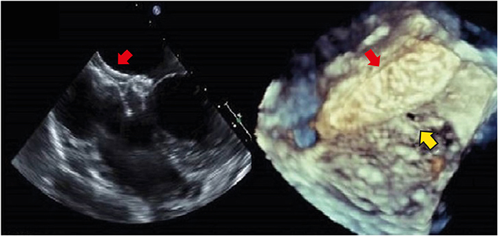
Figure 1 Transesophageal echo showed the atrial septal occluder attached to the anterior mitral leaflet. Yellow arrow indicates the perforated anterior mitral leaflet. Red arrow indicates atrial septal defect closure device.
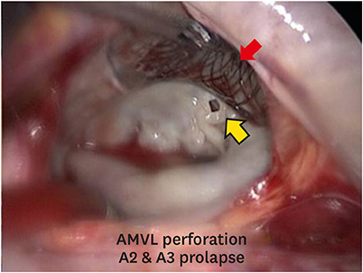
Figure 2 Mitral valve: anterior mitral leaflet perforation (about 3 mm) with diffuse anterior leaflet prolapsed. Amplatzer device was explanted including the adherent interatrial septum and the septal defect was reconstructed using Gore-Tex cardiovascular patch. Yellow arrow indicates the perforated anterior mitral leaflet. Red arrow indicates atrial septal defect closure device.
Reference
-
1. Moore J, Hegde S, El-Said H, et al. Transcatheter device closure of atrial septal defects: a safety review. JACC Cardiovasc Interv. 2013; 6:433–442.2. Masura J, Gavora P, Podnar T. Long-term outcome of transcatheter secundum-type atrial septal defect closure using Amplatzer septal occluders. J Am Coll Cardiol. 2005; 45:505–507.
Article3. Mishaly D, Ghosh P, Preisman S. Minimally invasive congenital cardiac surgery through right anterior minithoracotomy approach. Ann Thorac Surg. 2008; 85:831–835.
Article
- Full Text Links
-
- Actions
-
Cited
- CITED
-
- Close
- Share
-
- Similar articles
-
- Transcatheter Closure of Secundum Atrial Septal Defect with the Amplatzer Septal Occluder
- The Operative Management of Embolized Septal Occluder at Ascending Aorta
- Late Migration of Amplatzer Septal Occluder Device to the Descending Thoracic Aorta
- A Case of Turner's Syndrome Associated with Atrial Septal Defect and Mitral Valve Prolapse
- Surgical Extraction of an Embolized Atrial Septal Defect Occluder Device into Pulmonary Artery after Percutaneous Closure
