Incidentally found cystic lymphangioma of the adrenal gland in an elderly male cadaver
- Affiliations
-
- 1Medical Course, Jeju National University School of Medicine, Jeju, Korea.
- 2Department of Pathology, Jeju National University School of Medicine, Jeju, Korea.
- 3Department of Anatomy, Jeju National University School of Medicine, Jeju, Korea. spyoon@jejunu.ac.kr
- 4Institute for Medical Science, Jeju National University, Jeju, Korea.
- KMID: 2460197
- DOI: http://doi.org/10.12701/yujm.2019.00262
Abstract
- Adrenal cystic lymphangiomas are extremely rare entities that are often identified incidentally, with less than 60 cases reported to date. We found a protruding ovoid mass consisting of a multiloculated cystic lesion within right adrenal gland in the cadaver of a 75-year-old Korean man. The epithelial cells lining the adrenal cyst were diffusely positive for cluster of differentiation 31 and podoplanin, and negative for pan-cytokeratin. The histopathological diagnosis confirmed a cystic lymphangioma arising from the adrenal gland. Post-mortem findings of the present case are discussed based on the clinicopathological features of adrenal cystic lymphangiomas.
Keyword
MeSH Terms
Figure
-
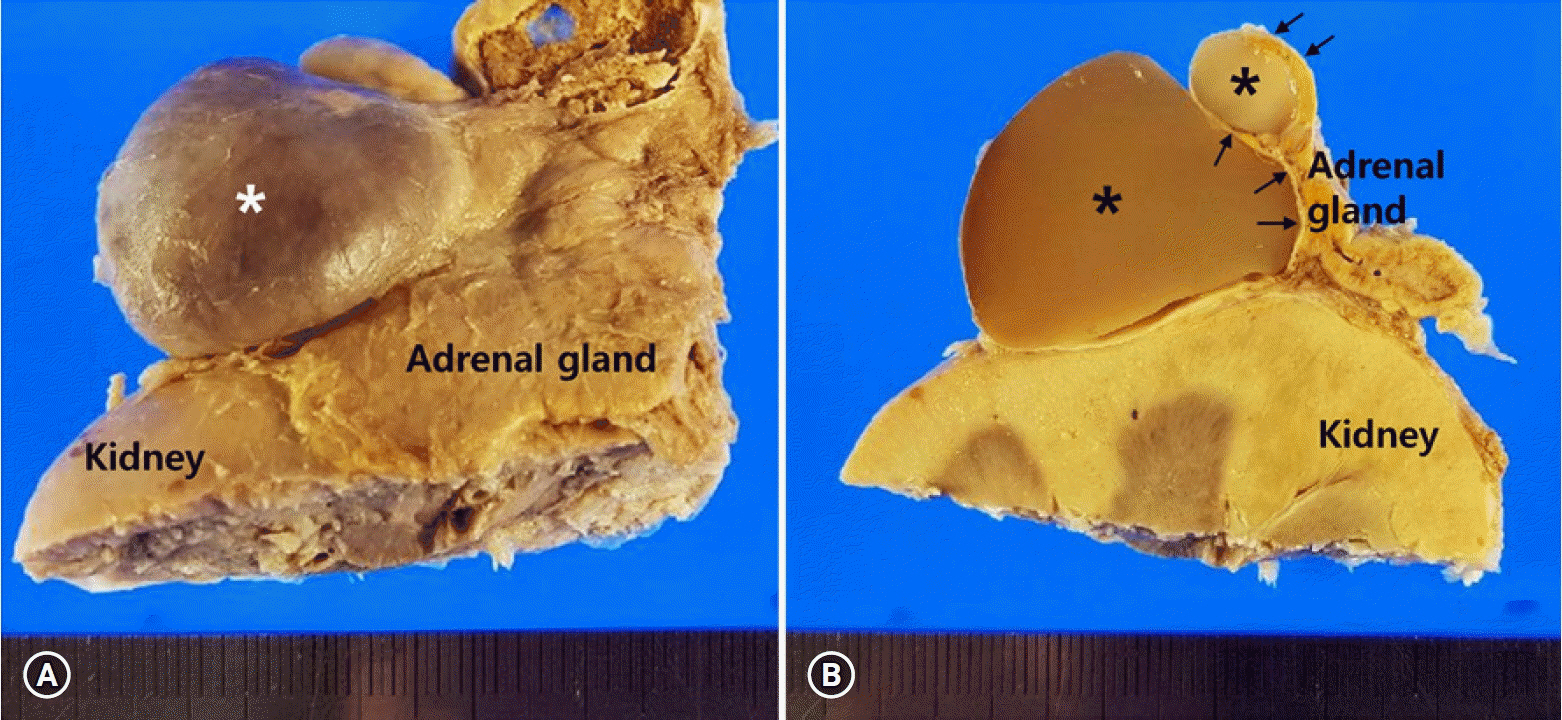
Fig. 1. Gross findings of the right-sided adrenal cystic mass. (A) The protruding mass (asterisk) shows smooth outer surface. (B) The multilocular cystic structures (asterisks) are surrounded by flattened adrenal parenchyma (arrows) on the section surface.

Fig. 2. Microscopic findings of the right-sided adrenal cystic lesion. (A) Multilocular cystic spaces are present. (B) Entrapped adrenal parenchyma (arrows) is present. (C) The cystic wall is lined by a single layer of flattened endothelial cells (arrowheads) (hematoxylin and eosin stain, ×40 [A and B] and ×200 [C]). The lining endothelial cells are positive for cluster of differentiation 31 (D) and podoplanin (E), and negative for pan-cytokeratin (F) (immunohistochemical stain, ×40 [D] and ×100 [E and F]).
Reference
-
References
1. Zheng W, Fung KM, Cheng L, Osunkoya AO. Benign vascular tumors, cysts, and pseudocysts of the adrenal gland: a contemporary multi-institutional clinicopathological analysis of 55 cases. Hum Pathol. 2018; 82:95–102.
Article2. Pollack HM. Miscellaneous conditions of the adrenals and adrenal pseudotumors. Clin Urogr. 1990; 3:2403–5.3. Rozenblit A, Morehouse HT, Amis ES Jr. Cystic adrenal lesions: CT features. Radiology. 1996; 201:541–8.
Article4. Tagge DU, Baron PL. Giant adrenal cyst: management and review of the literature. Am Surg. 1997; 63:744–6.5. Foster DG. Adrenal cysts. Review of literature and report of case. Arch Surg. 1966; 92:131–43.6. Longo JM, Jafri SZ, Bis KB. Adrenal lymphangioma: a case report. Clin Imaging. 2000; 24:104–6.
Article7. Rowe SP, Bishop JA, Prescott JD, Salvatori R, Fishman EK. CT appearance of adrenal cystic lymphangioma: radiologic-pathologic correlation. AJR Am J Roentgenol. 2016; 206:81–5.
Article8. Ates LE, Kapran Y, Erbil Y, Barbaros U, Dizdaroglu F. Cystic lymphangioma of the right adrenal gland. Pathol Oncol Res. 2005; 11:242–4.
Article9. Kafadar MT, Özyuvalı E, Miryaguboğlu AM, Çaviş T, İnan A. Incidental giant adrenal lymphangioma presenting as nonfunctional cystic mass. Turk J Surg. 2018; 1–3. [Epub ahead of print].
Article10. Michalopoulos N, Laskou S, Karayannopoulou G, Pavlidis L, Kanellos I. Adrenal gland lymphangiomas. Indian J Surg. 2015; 77(Suppl 3):1334–42.
Article11. Poncelet V. Retroperitoneal cystic lymphangioma. J Belge Radiol. 1998; 81:245.12. Ellis CL, Banerjee P, Carney E, Sharma R, Netto GJ. Adrenal lymphangioma: clinicopathologic and immunohistochemical characteristics of a rare lesion. Hum Pathol. 2011; 42:1013–8.
Article13. Bibi M, Sellami A, Taktak T, Chelly B, Ghorbel Z, Zouari H, et al. Giant cystic lymphangioma of adrenal gland: a case report and review of the literature. Urol Case Rep. 2018; 22:6–7.
Article14. Kim KH, Lee JI, Bae JM. Significant growth of adrenal lymphangioma: a case report and review of the literature. Int J Surg Case Rep. 2015; 17:48–50.
Article15. Tanuma Y, Kimura M, Sakai S. Adrenal cyst: a review of the Japanese literature and report of a case. Int J Urol. 2001; 8:500–3.
Article16. Satou T, Uesugi T, Nakai Y, Hayashi Y, Imano M, Hashimoto S. Case of adrenal lymphangioma with atypical lymphocytes in aspirate cytology. Diagn Cytopathol. 2003; 29:87–90.
Article
