Anti-oxidative Effect of Piperine from Piper nigrum L. in Caenorhabditis elegans
- Affiliations
-
- 1College of Pharmacy, Woosuk University, Jeonju 55338, Republic of Korea. dkkim@woosuk.ac.kr
- KMID: 2459969
- DOI: http://doi.org/10.20307/nps.2019.25.3.255
Abstract
- Piper nigrum L. (Piperaceae), which is a well-known food seasoning, has been used as a traditional medicine for the treatment of vomiting, abdominal pain, diarrhea and anorexia in Korea, China and Japan. Methanol extract from the fruit of P. nigrum was successively partitioned as n-hexane, methylene chloride, ethyl acetate, n-butanol and Hâ‚‚O soluble fractions. Among those fractions the ethyl acetate soluble fraction showed the most potent DPPH radical scavenging activity, and piperine was isolated from the ethyl acetate fraction. To know the antioxidant activity of piperine, we tested the activities of superoxide dismutase (SOD) and catalase together with oxidative stress tolerance and intracellular ROS level in Caenorhabditis elegans. To investigate whether piperine-mediated increased stress tolerance was due to regulation of stress-response gene, we quantified SOD-3 expression using transgenic strain including CF1553. Consequently, piperine enhanced SOD and catalase activities of C. elegans, and reduced intracellular ROS accumulation in a dose-dependent manner. Moreover, piperine-treated CF1553 worms exhibited significantly higher SOD-3::GFP intensity.
MeSH Terms
Figure
-

Fig. 1 Structure of piperine.
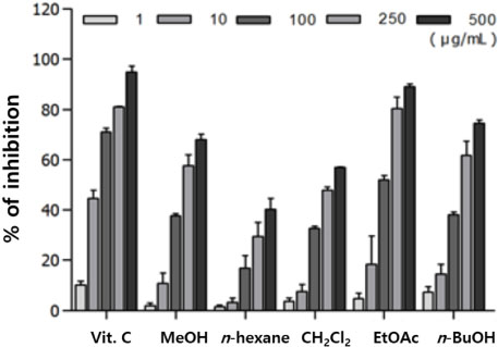
Fig. 2 DPPH radical scavenging effects of the MeOH extract and its subsequent fractions from Piper nigrum L.

Fig. 3 Oxidative stress tolerance of piperine was determined after the nematodes were transferred to 96-well plates containing 1 mM juglone. Statistical differences between the curves were analyzed by the log-rank test. 4-HBA (4-hydroxybenzoic acid): positive control.
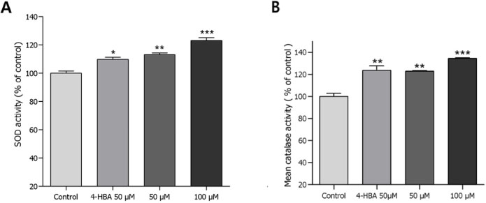
Fig. 4 Effects of piperine on the antioxidant enzyme activity of wild type N2 nematodes. (A) SOD activity was shown as a percentage of superoxide scavenged per control. (B) Catalase activity was expressed as a percentage of decrease in residual H2O2 measured by a spectrophotometric method. Differences compared with the control were considered significant at *p < 0.05, **p < 0.01, and ***p < 0.001 by the one-way ANOVA. 4-HBA (4-hydroxybenzoic acid): positive control.
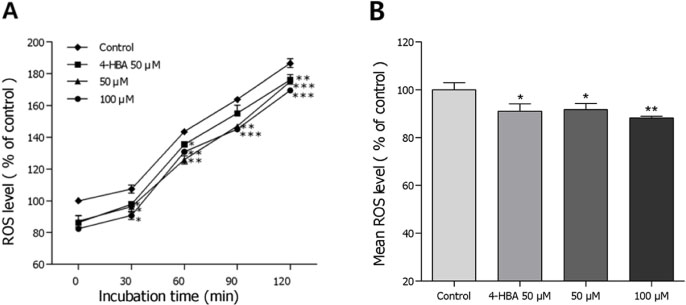
Fig. 5 Effects of piperine on the intracellular ROS levels of wild-type N2 nematodes. Intracellular ROS accumulation was examined in a microplate fluorescence reader at 535 nm (emission) and 485 nm (excitation). (A) Plates were read for 120 min. (B) The average percentages of intracellular ROS accumulation were presented. Differences compared with the control were considered significant at *p < 0.05, **p < 0.01, and ***p < 0.001 by the one-way ANOVA. 4-HBA (4-hydroxybenzoic acid): positive control.
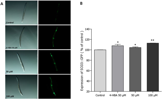
Fig. 6 Effects of piperine on the expression of SOD-3 (CF1553) was determined using transgenic nematodes. (A) Images of SOD-3::GFP expressions of CF1553 nematodes in the presence or absence of piperine. (B) The mean GFP-expressing intensity of CF1553 mutants was expressed as mean ± S.E.M. of values from 90 worms per each experiment. Data are expressed as the mean ± standard deviation of three independent experiments (N=3). Differences compared with the control were considered significant at *p < 0.05 and **p < 0.01 by one-way ANOVA. 4-HBA (4-hydroxybenzoic acid): positive control.
Reference
-
1. Parveen A, Akash MS, Rehman K, Kyunn WW. Crit Rev Eukaryot Gene Expr. 2016; 26:143–160.2. Prasad S, Gupta SC, Tyagi AK. Cancer Lett. 2017; 387:95–105.3. Pizzino G, Irrera N, Cucinotta M, Pallio G, Mannino F, Arcoraci V, Squadrito F, Altavilla D, Bitto A. Oxid Med Cell Longev. 2017; 2017:8416763.4. Partridge L, Gems D. Nat Rev Genet. 2002; 3:165–175.5. Weinert BT, Timiras PS. J Appl Physiol. 2003; 95:1706–1716.6. Deng Y, Sriwiriyajan S, Tedasen A, Hiransai P, Graidist P. J Ethnopharmacol. 2016; 188:87–95.7. Tasleem F, Azhar I, Ali SN, Perveen S, Mahmood ZA. Asian Pac J Trop Med. 2014; 7S1:S461–S468.8. Nikolić MM, Jovanović KK, Marković TL, Marković DL, Gligorijević NN, Radulović SS, Kostić M, Glamoćlija JM, Soković MD. J Pharm Pharmacol. 2017; 69:1606–1614.9. Ternes W, Krause EL. Anal Bioanal Chem. 2002; 374:155–160.
Article10. Brenner S. Genetics. 1974; 77:71–94.11. Mekheimer RA, Sayed AA, Ahmed EA. J Med Chem. 2012; 55:4169–4177.12. Aebi H. Methods Enzymol. 1984; 105:121–126.13. Kim HN, Seo HW, Kim BS, Lim HJ, Lee HN, Park JS, Yoon YJ, Oh JW, Oh MJ, Kwon J, Oh CH, Cha DS, Jeon H. Nat Prod Sci. 2015; 21:128–133.
Article14. Seo HW, Cheon SM, Lee MH, Kim HJ, Jeon H, Cha DS. Evid Based Complement Alternat Med. 2015; 2015:524878.15. Waters JC. J Cell Biol. 2009; 185:1135–1148.16. Kudryavtseva AV, Krasnov GS, Dmitriev AA, Alekseev BY, Kardymon OL, Sadritdinova AF, Fedorova MS, Pokrovsky AV, Melnikova NV, Kaprin AD, Moskalev AA, Snezhkina AV. Oncotarget. 2016; 7:44879–44905.17. Surco-Laos F, Dueñas M, González-Manzano S, Cabello J, Santos-Buelga C, González-Paramás AM. Food Res Int. 2012; 46:514–521.18. Liao CY, Kennedy BK. Cell Res. 2016; 26:143–144.19. Maurya PK, Noto C, Rizzo LB, Rios AC, Nunes SO, Barbosa DS, Sethi S, Zeni M, Mansur RB, Maes M, Brietzke E. Prog Neuropsychopharmacol Biol Psychiatry. 2016; 65:134–144.20. Heink AE, Parrish AN, Thorgaard GH, Carter PA. Aquat Toxicol. 2013; 144-145:75–82.21. Homayouni-Tabrizi M, Asoodeh A, Soltani M. J Food Drug Anal. 2017; 25:567–575.22. Mittal R, Gupta RL. Methods Find Exp Clin Pharmacol. 2000; 22:271–274.23. Verma N, Bal S, Gupta R, Aggarwal N, Yadav A. J Diet Suppl. 2018; 1–12.24. Umar S, Golam Sarwar AH, Umar K, Ahmad N, Sajad M, Ahmad S, Katiyar CK, Khan HA. Cell Immunol. 2013; 284:51–59.
- Full Text Links
-
- Actions
-
Cited
- CITED
-
- Close
- Share
-
- Similar articles
-
- Acanthopanax sessiliflorus stem confers increased resistance to environmental stresses and lifespan extension in Caenorhabditis elegans
- Effect of aspect ratio on the uptake and toxicity of hydroxylated-multi walled carbon nanotubes in the nematode, Caenorhabditis elegans
- Comparative study of oxidative stress caused by anthracene and alkyl-anthracenes in Caenorhabditis elegans
- Involvement of Caenohabditis elegans MAPK Signaling Pathways in Oxidative Stress Response Induced by Silver Nanoparticles Exposure
- The Longevity Properties of 1,2,3,4,6-Penta-O-Galloyl-beta-D-Glucose from Curcuma longa in Caenorhabditis elegans
