Bone Involvement of Diffuse Large B Cell Lymphoma (DLBCL) Showing Unusual Manifestations Mimicking Chronic Osteomyelitis in a 58-Year-Old Man: Case Report and Clinical Application of Diffusion Weighted Magnetic Resonance Imaging
- Affiliations
-
- 1Department of Radiology, Jeju National University Hospital, Jeju-si, Korea. we1977@naver.com
- 2Department of Pathology, Jeju National University Hospital, Jeju-si, Korea.
- KMID: 2459882
- DOI: http://doi.org/10.13104/imri.2019.23.3.270
Abstract
- This study presents a case of diffuse large B cell lymphoma (DLBCL) in a 58-year-old man showing unusual manifestations mimicking chronic osteomyelitis. In this case review, we describe the imaging findings of DLBCL which mimics chronic osteomyelitis and review existing reports regarding the differential diagnosis of bone involvement of lymphoma and osteomyelitis through imaging and laboratory findings and diffusion-weighted magnetic resonance imaging (DWI) such as the advanced MRI sequence.
MeSH Terms
Figure
-
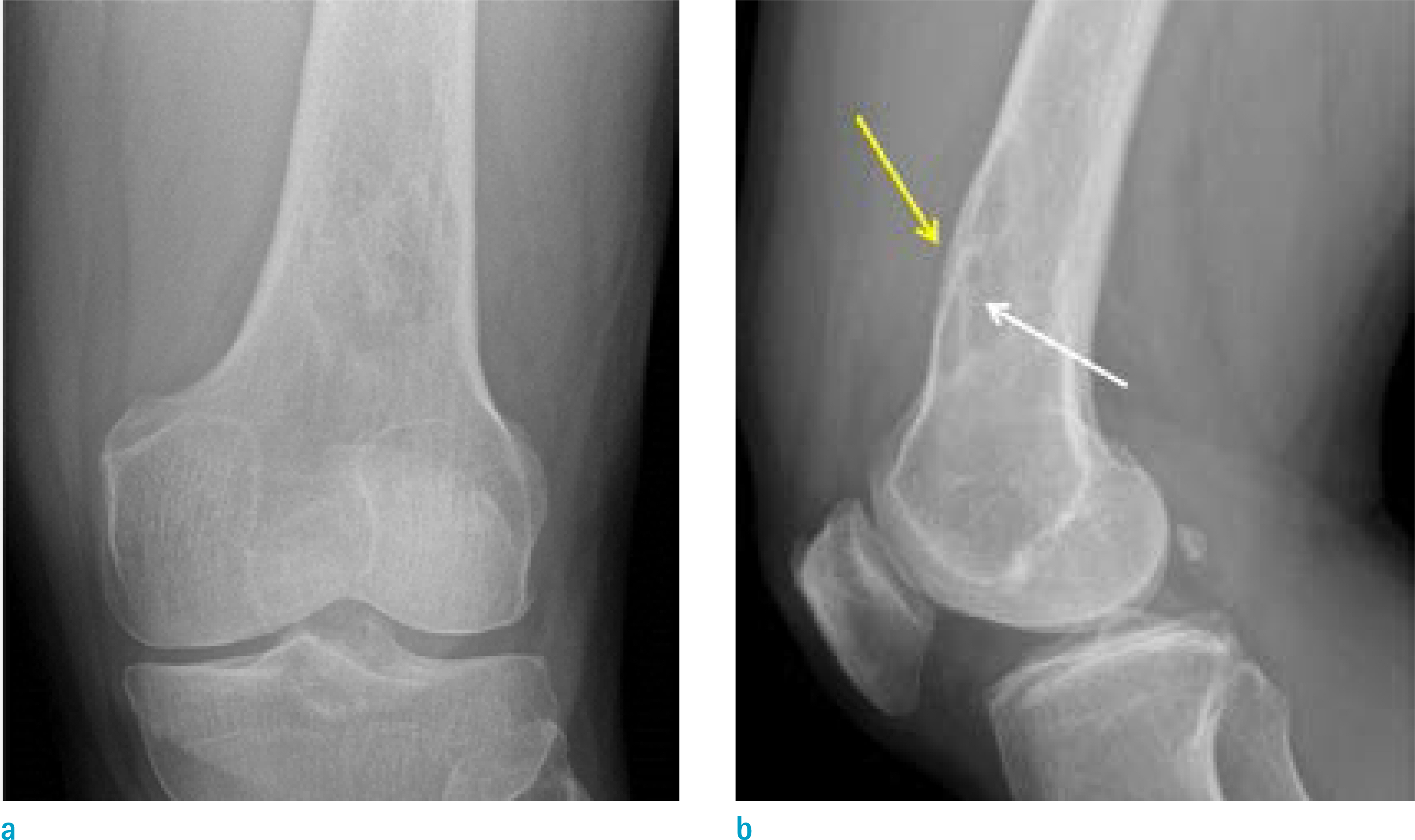
Fig. 1. AP (a) and lateral (b) radiographs of the left knee shows an osteolytic lesion with moth-eaten bone destruction in the distal diaphysis of the left femur. The lateral view of the left knee shows suspected sequestrum (white arrow) in the lesion and a single layer of periosteal new bone formation (yellow arrow) adjacent to the lesion.
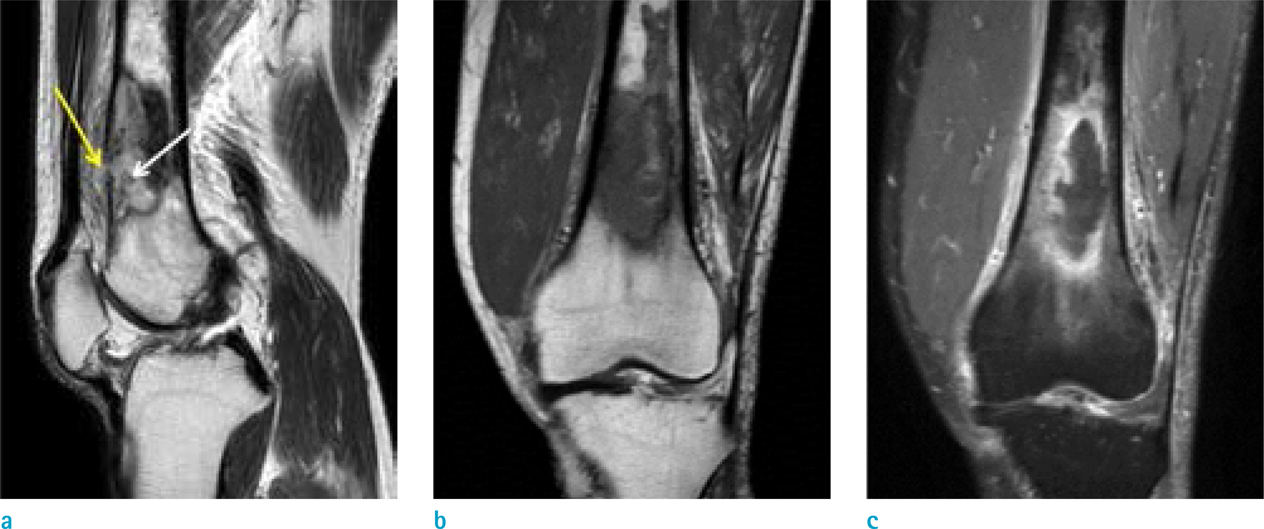
Fig. 2. Sagittal T2-weighted MR image (a) shows cortical perforation suggesting cloaca (yellow arrow) and suspected bony sequestrum (white arrow) in the distal diaphysis of the left femur. On coronal T1-weighted MR image (b), the lesion of the left distal femur shows low signal intensity and on coronal contrast-enhanced fat-suppressed T1-weighted MR image (c), the lesion consisting of non-enhancing portion that has suspected necrosis and enhancing portion in the periphery of the lesion.
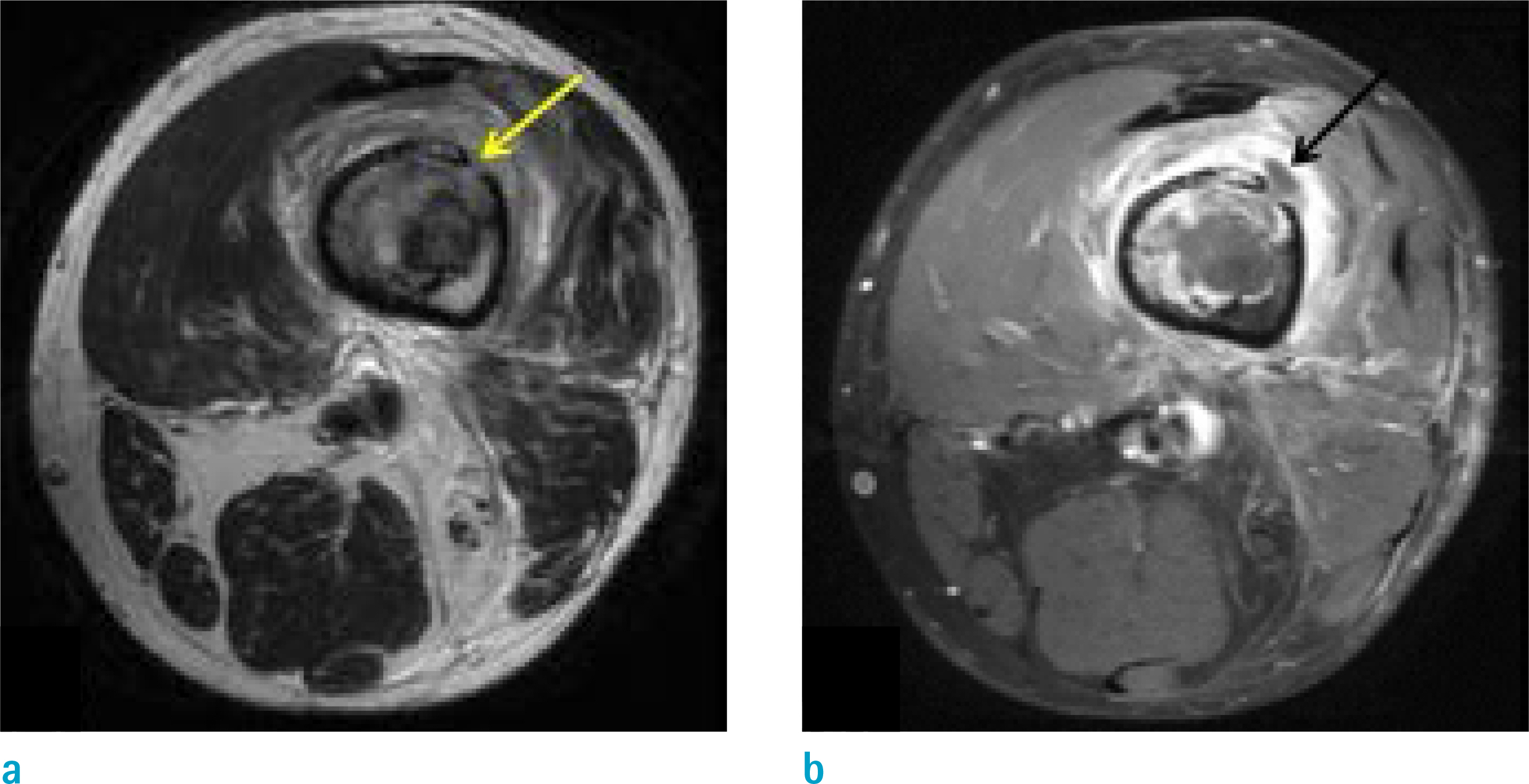
Fig. 3. Axial T2-weighted MR image (a) shows cortical perforation suggesting cloaca (yellow arrow) and axial contrast-enhanced fat-suppressed T1-weighted MR image (b) demonstrates the drainage of necrotic material out of the bone (black arrow).
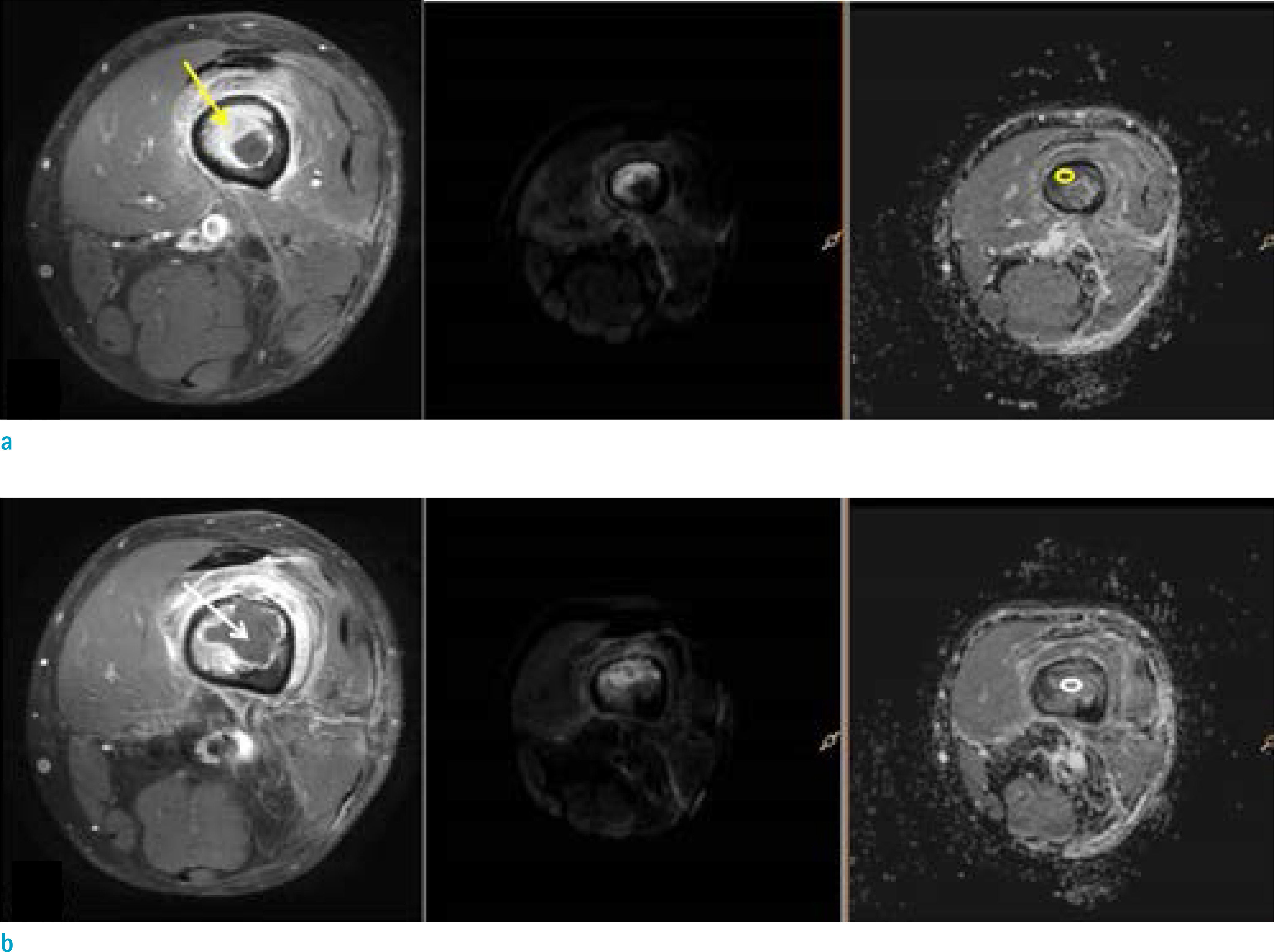
Fig. 4. The measurement of ADC value at b 800 s/mm2 using axial contrast-enhanced fat-suppressed T1-weighted MR images, diffusion weighted images (DWI) and apparent diffusion coefficient (ADC) map. (a) The mean ADC value of 0.84 × 10⁻³ mm2/s (b = 800 s/mm2) was found in the enhancing portion of the lesion (yellow arrow, yellow region of interest). (b) The mean ADC value was 1.87 × 10⁻³ mm2/s (b = 800 s/mm2) for the non-enhancing portion of the lesion (white arrow, white region of interest).

Fig. 5. Photomicrograph (a, Hematoxylin & Eosin [H&E], × 40) showing curetted tumor tissue admixed with bony trabeculae (arrows) and photomicrograph (b, H&E, × 40) showing necrotic areas (arrowheads). Photomicrograph (c, H&E, × 400) demonstrates neoplastic large lymphocytes and photomicrograph (d, × 200) illustrate that they are positive for CD20.

Fig. 6. PET CT demonstrates increased uptake in the left distal femur and lymph nodes of left inguinal, femoral, iliac, and paraaortic areas.
Reference
-
References
1. Kumar V, Abbas A, Fausto N, Aster J. Robbins and cotran pathologic basis of disease. 8th ed. Philadelphia, PA: Saunders/Elsevier. 2010. 606–607.2. Krishnan A, Shirkhoda A, Tehranzadeh J, Armin AR, Irwin R, Les K. Primary bone lymphoma: radiographic-MR imaging correlation. Radiographics. 2003; 23:1371–1383. discussion 1384–1377.
Article3. Jennin F, Bousson V, Parlier C, Jomaah N, Khanine V, Laredo JD. Bony sequestrum: a radiologic review. Skeletal Radiol. 2011; 40:963–975.
Article4. Markanday A. Acute phase reactants in infections: evidence-based review and a guide for clinicians. Open Forum Infect Dis. 2015; 2:ofv098.
Article5. Trichopoulos D, Psaltopoulou T, Orfanos P, Trichopoulou A, Boffetta P. Plasma C-reactive protein and risk of cancer: a prospective study from Greece. Cancer Epidemiol Biomarkers Prev. 2006; 15:381–384.
Article6. Wu S, Zhou Y, Hua HY, et al. Inflammation marker ESR is effective in predicting outcome of diffuse large B-cell lymphoma. BMC Cancer. 2018; 18:997.
Article7. Filograna L, Magarelli N, Cellini F, et al. Diffusion weighted imaging (DWI) and apparent diffusion coefficient (ADC) values for detection of malignant vertebral bone marrow lesions. Eur Rev Med Pharmacol Sci. 2018; 22:590–597.8. Wu X, Pertovaara H, Dastidar P, et al. ADC measurements in diffuse large B-cell lymphoma and follicular lymphoma: a DWI and cellularity study. Eur J Radiol. 2013; 82:e158–164.
Article9. Ozgen B, Oguz KK, Cila A. Diffusion MR imaging features of skull base osteomyelitis compared with skull base malignancy. AJNR Am J Neuroradiol. 2011; 32:179–184.
Article10. Abdel Razek AAK, Samir S. Diagnostic performance of diffusion-weighted MR imaging in differentiation of diabetic osteoarthropathy and osteomyelitis in diabetic foot. Eur J Radiol. 2017; 89:221–225.
Article
- Full Text Links
-
- Actions
-
Cited
- CITED
-
- Close
- Share
-
- Similar articles
-
- Multifocal Extranodal Involvement in Diffuse Large B-cell Lymphoma (DLBCL): Bone, Soft Tissue, Testis, and Kidney
- Diffuse Large B-Cell Lymphoma Associated with a Chronic Inflammatory Condition Induced by Metallic Implants: A Case Report
- MR Imaging Features of Primary Cutaneous Diffuse Large B-cell Lymphoma: A Case Report
- A Case of Diffuse Large B Cell Lymphoma in Middle Ear Cavity
- Primary Bone Lymphoma of the Distal Tibia Mimicking Brodie's Abscess
