Primary Rhabdomyosarcoma of the Breast: Study of Three Cases at One Institution with a Review of Primary Breast Sarcomas
- Affiliations
-
- 1Department of Pathology, Asan Medical Center, University of Ulsan College of Medicine, Seoul, Korea. kjc@amc.seoul.kr
- 2Department of Surgery, Asan Medical Center, University of Ulsan College of Medicine, Seoul, Korea.
- 3Department of Pediatric Surgery, Asan Medical Center, University of Ulsan College of Medicine, Seoul, Korea.
- KMID: 2459579
- DOI: http://doi.org/10.4132/jptm.2019.07.22
Abstract
- BACKGROUND
Primary breast sarcoma (PBS) is rare, comprising approximately 1% of breast malignancies. Rhabdomyosarcoma (RMS) accounts for an extremely small proportion of PBSs, often leading to delayed histologic confirmation.
METHODS
Upon reviewing Asan Medical Center's pathology database between 2000 and 2018, 41 PBS cases were retrieved, including three cases of primary RMS of the breast. Their clinicopathological features were analyzed, and the literature related to PBS and primary RMS of the breast was reviewed.
RESULTS
We identified three primary breast RMS cases from our institution database, comprising 7.3% of PBS: one case each of spindle cell/sclerosing RMS (ssRMS), alveolar RMS (aRMS), and embryonal RMS (eRMS). All cases involved adolescents or young adults (14, 16, and 25 years, respectively) who underwent mastectomy or radiotherapy and were confirmed using immunohistochemical testing for myogenin, desmin, and myogenic differentiation. The ssRMS patient experienced recurrence at the operation site 4 months post-surgery despite undergoing concurrent chemoradiotherapy. The aRMS patient had multiple metastases at diagnosis and showed FAX3-FOXO1 fusion transcripts; she died 22 months after the diagnosis. The eRMS patient had enlarged axillary lymph nodes; post-radiotherapy, the lesion recurred as multiple metastases to the bone and lung. She died 18 months post-diagnosis.
CONCLUSIONS
Our experience on RMS cases suggests that spindle cell or small round cell malignancy in breasts of young female should raise suspicion for the possibility of primary or secondary RMS. To our knowledge, this is the second report of primary breast ssRMS and it may help clinicians who encounter this rare disease in the future.
MeSH Terms
Figure
-

Fig. 1. Primary breast sarcoma of patient A. (A) A 12×9.5×8.3-cm-sized heterogeneously enhancing mass in the left breast (yellow arrow). (B) Grayish-yellow cut surface of the mass with multifocal hemorrhagic spots and central cavity formation. (C, D) Spindle cells exhibiting a fascicular or storiform growth pattern with a small amount of myxoid matrix. The image showing highly pleomorphic elongated nuclei and pale to eosinophilic cytoplasm, with high mitotic rates (up to 94/10 high power fields). (E–H) Immunohistochemical staining showing positivity of tumor cells for MyoD1 (E), desmin (F), and myogenin (G) and negativity for MNF-116 (H).
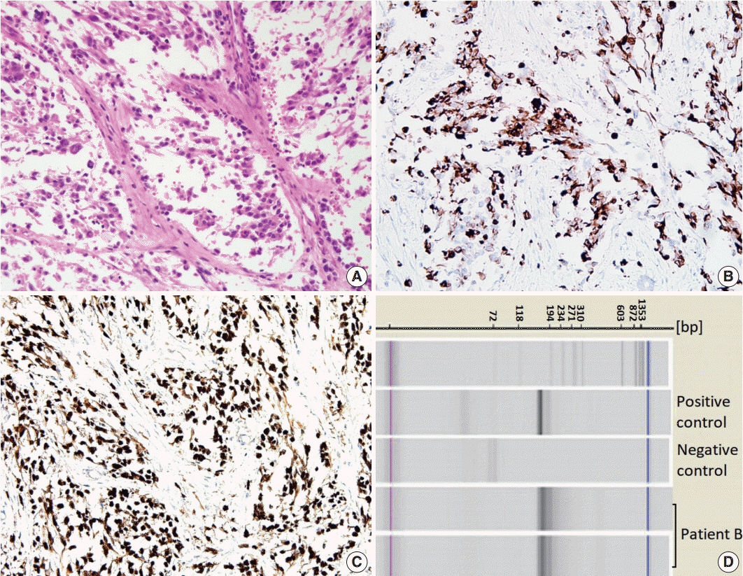
Fig. 2. Primary breast sarcoma of patient B. (A) Primitive round-to-polygonal cells comprising the nests separated by fibrous septa and showing a loss of cellular cohesion. (B, C) Immunohistochemical staining for desmin (B) and myogenin (C). (D) PAX3-FKHR (FOXO1) fusion identified by PAX-3/PAX-7-FKHR (FOXO1) nested reverse transcription polymerase chain reaction analysis.

Fig. 3. Primary breast sarcoma of patient C. (A) Magnetic resonance imaging of the breast showing multifocal, infiltrating, ill-defined, enhancing masses in the left lower inner, upper inner, and lower outer quadrants. (B) Hyperchromatic small round cells with scanty cytoplasm exhibiting solid-sheet and cord-like growth patterns. (C, D) Immunohistochemical staining showing negativity for desmin (C) and positivity for myogenin (D).

Fig. 4. Age distribution of primary breast sarcoma (PBS) according to subtype. Angiosarcoma was the most common sarcoma, followed by extra-skeletal osteosarcoma, liposarcoma, and dermatofibrosarcoma protuberans (DFSP). The most prevalent age-group was 50–60 years. PBS was rare in people aged less than 30, and rhabdomyosarcoma was the most common subtype in this age-group. UPS, undifferentiated pleomorphic sarcoma; MPNST, malignant peripheral nerve sheath tumor.
Reference
-
1. Lim SZ, Ong KW, Tan BK, Selvarajan S, Tan PH. Sarcoma of the breast: an update on a rare entity. J Clin Pathol. 2016; 69:373–81.
Article2. Hicks DG, Lester SC. Diagnostic pathology: breast. Salt Lake City: Saunders-Elsevier;2016. p. 496–577. 2nd.3. Chugh R, Baker L. Nonepithelial malignancies of the breast. Oncology (Williston Park). 2004; 18:665–73.4. Yin M, Mackley HB, Drabick JJ, Harvey HA. Primary female breast sarcoma: clinicopathological features, treatment and prognosis. Sci Rep. 2016; 6:31497.
Article5. Attili VS, Dadhich HK, Ramarao C, Bapsy PP, Ramachandra C, Anupama G. A case of primary rhabdomyosarcoma of the breast. Indian J Surg. 2007; 69:201–2.
Article6. Kebudi R, Koc BS, Gorgun O, Celik A, Kebudi A, Darendeliler E. Breast metastases in children and adolescents with rhabdomyosarcoma: a large single-institution experience and literature review. J Pediatr Hematol Oncol. 2017; 39:67–71.
Article7. Audino AN, Setty BA, Yeager ND. Rhabdomyosarcoma of the breast in adolescent and young adult (AYA) women. J Pediatr Hematol Oncol. 2017; 39:62–6.
Article8. Fletcher CD, Bridge JA, Hogendoorn P, Mertens F. WHO classification of tumors of soft tissue and bone. Lyon: International Agency for Research on Cancer;2013. p. 127–36. 4th.9. Rasinariu A, Andreiuolo F, Terrier P, Balleyguier C, Delaloge S, Vielh P. Primary spindle rhabdomyosarcoma of the breast in an adult female. Cytopathology. 2011; 22:137–9.
Article10. Kim DY, Seol YM, Kim H, Kim A, Choi YJ. Primary rhabdomyosarcoma of the breast in a 17-year-old girl: case report. Medicine (Baltimore). 2017; 96:e9076.11. Bland KI, Klimberg VS, Copeland E, Gradishar WJ. The breast: comprehensive management of benign and malignant disease. Philadelphia: Saunders-Elsevier;2018. p. 156–68. 5th.12. Li N, Cusido MT, Navarro B, et al. Breast sarcoma: a case report and review of literature. Int J Surg Case Rep. 2016; 24:203–5.
Article13. Schnitt SJ, Collins LC. Biopsy interpretation of the breast. 2nd. Philadelphia: Lippincott Williams & Wilkins;2013. p. 416–7.14. Italiano A, Largillier R, Peyrottes I, Hannoun-Levy JM, Lallement M, Thyss A. Primary embryonal rhabdomyosarcoma of the breast in an adult female. Breast J. 2005; 11:214.
Article15. Guerrero MA, Ballard BR, Grau AM. Malignant phyllodes tumor of the breast: review of the literature and case report of stromal overgrowth. Surg Oncol. 2003; 12:27–37.
Article16. Parham DM, Ellison DA. Rhabdomyosarcomas in adults and children: an update. Arch Pathol Lab Med. 2006; 130:1454–65.
Article17. Parham DM, Barr FG. Classification of rhabdomyosarcoma and its molecular basis. Adv Anat Pathol. 2013; 20:387–97.
Article18. Hays DM, Donaldson SS, Shimada H, et al. Primary and metastatic rhabdomyosarcoma in the breast: neoplasms of adolescent females, a report from the Intergroup Rhabdomyosarcoma Study. Med Pediatr Oncol. 1997; 29:181–9.
Article19. Herrera LJ, Lugo-Vicente H. Primary embryonal rhabdomyosarcoma of the breast in an adolescent female: a case report. J Pediatr Surg. 1998; 33:1582–4.
Article20. Binokay F, Soyupak SK, Inal M, Celiktas M, Akgul E, Aksungur E. Primary and metastatic rhabdomyosarcoma in the breast: report of two pediatric cases. Eur J Radiol. 2003; 48:282–4.
Article21. Vishnevskaia Ia V, Sharoev TA, Stepanova EV, Osipova LV. Rhabdomyosarcoma of the breast in girls. Arkh Patol. 2004; 66:47–51.22. Dausse F, Balu-Maestro C, Chapellier C, Leblanc-Talent P. Rhabdomyosarcoma of the breast. Clin Imaging. 2005; 29:337–41.
Article23. Nogi H, Kobayashi T, Kawase K, et al. Primary rhabdomyosarcoma of the breast in a 13-year-old girl: report of a case. Surg Today. 2007; 37:38–42.
Article24. Valera ET, Brassesco MS, Muglia VF, Scrideli CA, Tone LG. Alveolar rhabdomyosarcoma with breast involvement. Chin Med J (Engl). 2013; 126:998.25. Bhosale SJ, Kshirsagar AY, Sulhyan SR, Sulhyan SR. Rhabdomyosarcoma of the breast: a rare malignancy. Am J Case Rep. 2013; 14:250–2.26. Mondal SK, Mandal PK, Adhikari A, Basak B. Primary pleomorphic rhabdomyosarcoma of breast: report of a rare neoplasm. J Res Med Sci. 2014; 19:1200–2.27. Kallianpur AA, Shukla NK, Deo SV, Khanna P, Durgapal P. Primary mammary rhabdomyosarcoma in a nineteen year old female: a case report and review of literature. Indian J Cancer. 2015; 52:295–6.
Article28. Pareekutty NM, Bhagat M, Vora T, Qureshi SS. Rhabdomyosarcoma of the breast: report of two cases with the review of literature. J Indian Assoc Pediatr Surg. 2016; 21:81–3.
Article29. Yuan Y, Hou J, Pan Y. Rhabdomyosarcoma of the breast: report of a rare malignancy. Cancer Biol Ther. 2017; 18:676–80.
Article30. Bayramoglu Z, Kebudi R, Yilmaz R, et al. Primary rhabdomyosarcoma of the breast: imaging findings and literature review. Breast Care (Basel). 2018; 13:293–7.
Article31. Jean-Louis CJ. Alveolar rhabdomyosarcoma of the breast in adolescent female. Am Surg. 2018; 84:e352–3.
Article
- Full Text Links
-
- Actions
-
Cited
- CITED
-
- Close
- Share
-
- Similar articles
-
- An Unusual Imaging Finding of Breast Metastasis from Rhabdomyosarcoma
- Primary Osteosarcoma of the Breast: A case report
- Primary Angiosarcoma of the Breast: A Case Report
- Common and Uncommon Conditions of Breast Disease in Children and Adolescents: A Pictorial Review
- A Case of Primary Bilateral Angiosarcoma of the Breast
