From papyrus leaves to bioprinting and virtual reality: history and innovation in anatomy
- Affiliations
-
- 1Division of Thoracic Surgery, David Geffen School of Medicine, UCLA, Los Angeles, CA, USA.
- 2Department of Molecular, Cell, and Developmental Biology, UCLA, Los Angeles, CA, USA.
- 3Division of Pulmonary and Critical Care Medicine, David Geffen School of Medicine, UCLA, Los Angeles, CA, USA. bhartipaul@gmail.com
- KMID: 2459536
- DOI: http://doi.org/10.5115/acb.18.213
Abstract
- The human quest to master the anatomy and physiology of living systems started as early as 1600 BC, with documents from the Greeks, Indians, and Romans presenting the earliest systematic studies and advances. Following the fall of the Roman Empire, the progress slowed until the Renaissance renewed scientific interest in anatomy and physiology, ushering in an era of spectacular advances. Alongside the discoveries of modern science, innovations in media such as printing, photography and color reproduction, improved the accuracy of communicating science. Techniques for noninvasively viewing the human body, such as magnetic resonance imaging, opened up new ways of exploring and understanding anatomy, physiology, and disease pathogenesis. Advances in three-dimensional (3D)-technologies, including computer graphics and animation are directly linked to many advances in medicine and surgery. Anatomy education has come a long way from papyrus leaf inscriptions to computerized 3D modeling, holographic representation, and virtual reality-based software. The future presents unlimited options for studying and understanding anatomy as Google glasses, bioprinting, virtual reality, and allied technologies transform the world into a classroom. This review summarizes the journey of mankind to master anatomy and physiology.
MeSH Terms
Figure
-

Fig. 1 Edwin Smith Papyrus. Recto Columns 8 and 9. Picture from the archives of U.S. National Library of Medicine, https://ceb.nlm.nih.gov/projects/ttp/.

Fig. 2 Drawing (c. 1507) by Leonardo da Vinci showing the principal organs, vasculature, and the female urinogenital system. Source: http://www.visi.com/~reuteler/leonardo.html{{pd}}Category:LeonardodaVinci.
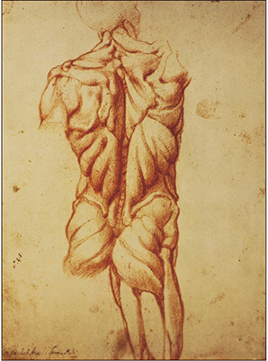
Fig. 3 An excerpt of Michelangelo's anatomical studies of the muscular system. Source: http://classicalpulse.blogspot.com/2013/06/michelangeloanatomy-studies.html.

Fig. 4 Vesalius drew accurate illustrations, with. Figures drawn in the artistic style of Renaissance realism. This figure of the musculoskeletal system stands in controposto. De Humani Corporis Fabrica. Basel, 1543. Woodcut. Picture from the archives of U.S. National Library of Medicine, https://www.nlm.nih.gov/dreamanatomy/da_g_I-B-1-07.html.
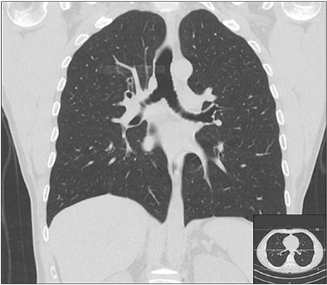
Fig. 5 A typical computed tomography scan through the coronal plane, showing the details of the cavity and parts of the abdomen. Right bottom: as seen through the Axial plane. A great improvement from century old paintings. Source: https://commons.wikimedia.org/w/index.php?title=Scrollable_highresolution_computed_tomography_images_of_a_normal_thorax&oldid=255941168.
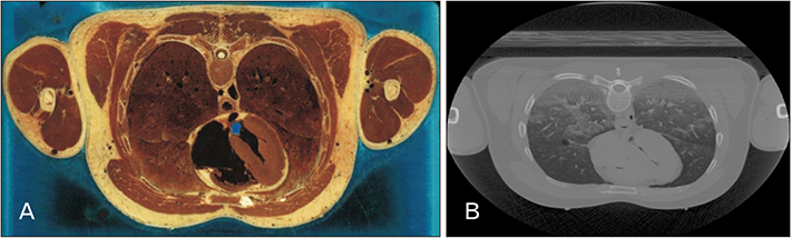
Fig. 6 This is from the thorax subset of the VHP project. (A) Photograph showing a section through Visible Human Male: thorax, including heart, lungs, spinal column, major vessels, musculature. (B) Computed tomography scan image of the same plane of the thorax. Source: from the archives of National Library of Medicine, https://www.nlm.nih.gov/research/visible/visible_gallery.html.

Fig. 7 Patient specific successive computed tomography (CT) scans are deconvoluted to make a 3D model. (A) Successive CT scans are placed back to back to create depth in an artistic way. (B–H) CT scan deconvolution. The 3D model created is rotated and shown from different angels and showing different layers, complexity of organ system. Multi-material FDM 3D printing can elucidate the skin and underlying organs. CT scan database: Lung Image Database Consortium image collection (LIDC).
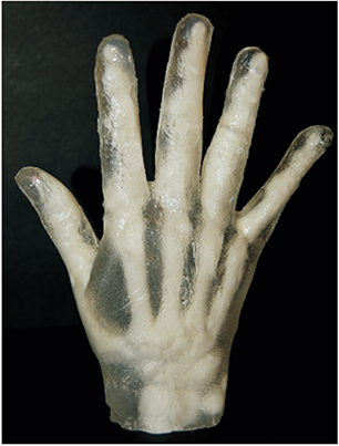
Fig. 8 3D printed model showing the skeletal structure (white) and the musculature (transparent). The template was made using a computed tomography bone scan and 3D hand scan and printed by dual extruder printing. Model organs can also be printed for students and practitioners.

Fig. 9 Jaw bone defect corrected with a bone mimic transplant made 3D printed from a computed tomography scan (shown by arrows).
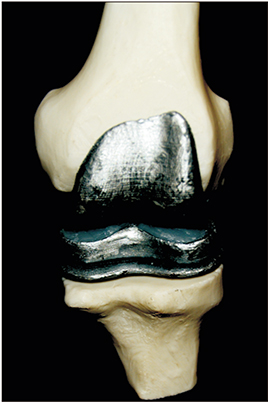
Fig. 10 Bone bio-engineering. Knee joint replacement or knee arthroplasty is commonly used to treat severe arthritis pain or injury. The prosthetic is 3D printed from a model based on the patient's computed tomography (CT) scan by metal laser sintering, CAD files: grab CAD. Alternatively, 3D printed bone scaffolds can be seeded with osteogenic cells derived from bone marrow stem cells or iPSCs and allowed to integrate normally. CT Database: Lung Image Database Consortium image collection.
Cited by 1 articles
-
The creation of a body donation program at Federal University of Juiz de Fora in Brazil: academic importance, challenges and donor profile
André Gustavo Fernandes de Oliveira, Augusto Ferreira Gonçalves, Júlia Nunes Soares, Letícia Henriques Neto Salgado, Bruno Silveira Santana, Matheus Venâncio Passos, Juliana Lopes de Oliveira Reis, Gustavo Candiá Arantes, Luís Filipe Sarmento Campos, Matheus Souza Carvalho, Lucas Cotrim Furtado da Gama, Alice Belleigoli Rezende
Anat Cell Biol. 2021;54(4):489-500. doi: 10.5115/acb.21.096.
Reference
-
1. Persaud TV, Loukas M, Tubbs RS. A history of human anatomy. Springfield, IL: Charles C Thomas Publisher Ltd.;2014.2. Aldersey-Williams H. Anatomies: a cultural history of the human body. New York: Norton & Company;2014.3. Vargas A, López M, Lillo C, Vargas MJ. The Edwin Smith papyrus in the history of medicine. Rev Med Chil. 2012; 140:1357–1362.4. Hallmann-MikoXMLLink_XYZajczak A. Ebers Papyrus. The book of medical knowledge of the 16th century B.C. Egyptians. Arch Hist Filoz Med. 2004; 67:5–14.5. Loukas M, Lanteri A, Ferrauiola J, Tubbs RS, Maharaja G, Shoja MM, Yadav A, Rao VC. Anatomy in ancient India: a focus on the Susruta Samhita. J Anat. 2010; 217:646–650.
Article6. Deepa B, Pushpalatha K. Contributions of Sushruta to anatomy. Anat J Afr. 2014; 3:362–365.7. Mahabir RC, Son-Hing J, Tam AL, Vastardis AD. Ancient Indian civilization: ahead by a nose. J Invest Surg. 2001; 14:3–5.8. Reverón RR. Herophilus and Erasistratus, pioneers of human anatomical dissection. Vesalius. 2014; 20:55–58.9. Bay NS, Bay BH. Greek anatomist herophilus: the father of anatomy. Anat Cell Biol. 2010; 43:280–283.
Article10. Fortuna S. The Latin editions of Galen's Opera omnia (1490-1625) and their prefaces. Early Sci Med. 2012; 17:391–412.
Article11. Trkalj G. Galen, macaques and the growth of the discipline of human anatomy. Am J Phys Anthropol. 2018; 165:277.12. Lascaratos J, Voros D, Tsiamis C. Paul of Aegina: landmark in surgical progress. World J Surg. 2003; 27:1336.13. Malomo AO, Idowu OE, Osuagwu FC. Lessons from history: human anatomy, from the origin to the Renaissance. Int J Morphol. 2006; 24:99–104.
Article14. Bertoloni Meli D. Early modern experimentation on live animals. J Hist Biol. 2013; 46:199–226.
Article15. Mavrodi A, Paraskevas G. Mondino de Luzzi: a luminous figure in the darkness of the Middle Ages. Croat Med J. 2014; 55:50–53.
Article16. Toledo-Pereyra LH. Leonardo da Vinci: the hidden father of modern anatomy. J Invest Surg. 2002; 15:247–249.
Article17. Zampieri F, ElMaghawry M, Zanatta A, Thiene G. Andreas vesalius: celebrating 500 years of dissecting nature. Glob Cardiol Sci Pract. 2015; 2015:66.
Article18. Van Hee R, Wells FC, Ballestriero R, Richardson R, Mazzarello P, Cani V, Catani M. The art of human anatomy: Renaissance to 21st century. Vesalius. 2014; 20:25–29.19. Pearson AS. Art and anatomy in Renaissance Italy: images from a scientific revolution. Sixteenth Century J. 2013; 44:601–602.20. DiDio LJ. Marcello Malpighi: the father of microscopic anatomy. Ital J Anat Embryol. 1995; 100 Suppl 1:3–9.21. Puccetti ML, Perugi L, Scarani P. Gaetano Giulio Zumbo. The founder of anatomic wax modeling. Pathol Annu. 1995; 30 Pt 2:269–281.22. Hajdu SI. A note from history: Rudolph Virchow, pathologist, armed revolutionist, politician, and anthropologist. Ann Clin Lab Sci. 2005; 35:203–205.23. Ghosh SK. Giovanni Battista Morgagni (1682–1771): father of pathologic anatomy and pioneer of modern medicine. Anat Sci Int. 2017; 92:305–312.
Article24. Doi K. Diagnostic imaging over the last 50 years: research and development in medical imaging science and technology. Phys Med Biol. 2006; 51:R5–R27.
Article25. Dong Z, Andrews T, Xie C, Yokoo T. Advances in MRI Techniques and Applications. Biomed Res Int. 2015; 2015:139043.
Article26. Fasel JH, Aguiar D, Kiss-Bodolay D, Montet X, Kalangos A, Stimec BV, Ratib O. Adapting anatomy teaching to surgical trends: a combination of classical dissection, medical imaging, and 3D-printing technologies. Surg Radiol Anat. 2016; 38:361–367.
Article27. Grignon B, Oldrini G, Walter F. Teaching medical anatomy: what is the role of imaging today? Surg Radiol Anat. 2016; 38:253–260.
Article28. Juanes JA, Prats A, Lagandara ML, Riesco JM. Application of the "Visible Human Project" in the field of anatomy: a review. Eur J Anat. 2003; 7:147–159.29. Spitzer VM, Whitlock DG. The Visible Human Dataset: the anatomical platform for human simulation. Anat Rec. 1998; 253:49–57.
Article30. Dai JX, Chung MS, Qu RM, Yuan L, Liu SW, Shin DS. The Visible Human Projects in Korea and China with improved images and diverse applications. Surg Radiol Anat. 2012; 34:527–534.
Article31. Zhang SX, Heng PA, Liu ZJ. Chinese visible human project. Clin Anat. 2006; 19:204–215.
Article32. Murgitroyd E, Madurska M, Gonzalez J, Watson A. 3D digital anatomy modelling: practical or pretty? Surgeon. 2015; 13:177–180.33. McMenamin PG, Quayle MR, McHenry CR, Adams JW. The production of anatomical teaching resources using three-dimensional (3D) printing technology. Anat Sci Educ. 2014; 7:479–486.
Article34. Chen B, Leng S, Yu L, Holmes D 3rd, Fletcher J, McCollough C. An open library of CT patient projection data. Proc SPIE Int Soc Opt Eng. 2016; 9783:97831B.
Article35. Zilverschoon M, Vincken KL, Bleys RL. The virtual dissecting room: Creating highly detailed anatomy models for educational purposes. J Biomed Inform. 2017; 65:58–75.
Article36. Fredieu JR, Kerbo J, Herron M, Klatte RS, Cooke M. Anatomical models: a digital revolution. Med Sci Educ. 2015; 25:183–194.
Article37. Ward TM, Wertz CI, Mickelsen W. Anatomage table enhances radiologic technology education. Radiol Technol. 2018; 89:304–306.38. Nicholson DT, Chalk C, Funnell WR, Daniel SJ. Can virtual reality improve anatomy education? A randomised controlled study of a computer-generated three-dimensional anatomical ear model. Med Educ. 2006; 40:1081–1087.
Article39. Olasky J, Sankaranarayanan G, Seymour NE, Magee JH, Enquobahrie A, Lin MC, Aggarwal R, Brunt LM, Schwaitzberg SD, Cao CG, De S, Jones DB. Identifying opportunities for virtual reality simulation in surgical education: a review of the Proceedings from the Innovation, Design, and Emerging Alliances in Surgery (IDEAS) Conference: VR Surgery. Surg Innov. 2015; 22:514–521.40. Devcic Z, Kesselman A, Shah R, AbdelRazek M, Kothary N. Augmented virtual reality assisted treatment planning for splenic artery aneurysms: a pilot study. J Vasc Interv Radiol. 2018; 29:S17.41. Armstrong AJ, Anand A, Edenbrandt L, Bondesson E, Bjartell A, Widmark A, Sternberg CN, Pili R, Tuvesson H, Nordle O, Carducci MA, Morris MJ. Phase 3 assessment of the automated bone scan index as a prognostic imaging biomarker of overall survival in men with metastatic castration-resistant prostate cancer: a secondary analysis of a randomized clinical trial. JAMA Oncol. 2018; 4:944–951.42. Li L, Yu F, Shi D, Shi J, Tian Z, Yang J, Wang X, Jiang Q. Application of virtual reality technology in clinical medicine. Am J Transl Res. 2017; 9:3867–3880.43. Yudkowsky R, Luciano C, Banerjee P, Schwartz A, Alaraj A, Lemole GM Jr, Charbel F, Smith K, Rizzi S, Byrne R, Bendok B, Frim D. Practice on an augmented reality/haptic simulator and library of virtual brains improves residents' ability to perform a ventriculostomy. Simul Healthc. 2013; 8:25–31.
Article44. Heller TB, Hill RM, Saggal AF. 3D Systems, Inc. Google patent WO1991012120A1. 1991. 08. 22.45. Ventola CL. Medical Applications for 3D printing: current and projected uses. P T. 2014; 39:704–711.46. Garcia J, Yang Z, Mongrain R, Leask RL, Lachapelle K. 3D printing materials and their use in medical education: a review of current technology and trends for the future. BMJ Simul Technol Enhanc Learn. 2018; 4:27–40.
Article47. Chia HN, Wu BM. Recent advances in 3D printing of biomaterials. J Biol Eng. 2015; 9:4.
Article48. Seol YJ, Kang HW, Lee SJ, Atala A, Yoo JJ. Bioprinting technology and its applications. Eur J Cardiothorac Surg. 2014; 46:342–348.
Article49. Dababneh AB, Ozbolat IT. Bioprinting technology: a current state-of-the-art review. J Manuf Sci Eng. 2014; 136:061016.
Article50. Morris S. Future of 3D printing: How 3D bioprinting technology can revolutionize healthcare? Birth Defects Res. 2018; 110:1098–1101.
Article51. Brazina D, Fojtik R, Rombova Z. 3D visualization in teaching anatomy. Procedia Soc Behav Sci. 2014; 143:367–371.
Article52. Melo JS, Brasil LM, Balaniuk R, Ferneda E, Santana JS. Intelligent tutoring systems based on ontologies and 3D visualization platforms in the teaching of the human anatomy. In : Dössel O, Schlegel WC, editors. World Congress on Medical Physics and Biomedical Engineering, 2009 Sep 7-12, Munich, Germany. IFMBE Proceedings, Vol. 25. Berli: Springer;2009. p. 16–17.53. Negi S, Dhiman S, Sharma RK. Basics and applications of rapid prototyping medical models. Rapid Prototyp J. 2014; 20:256–267.
Article54. Carter Y, Allard TT, Moore NC, Goertzen AL, Klonisch T, Hoppa RD. The role of 3D printing in teaching and education in human skeletal anatomy. FASEB J. 2009; 23:1 Suppl. 479.55. Patuzzo S, Goracci G, Gasperini L, Ciliberti R. 3D bioprinting technology: scientific aspects and ethical issues. Sci Eng Ethics. 2018; 24:335–348.
Article
- Full Text Links
-
- Actions
-
Cited
- CITED
-
- Close
- Share
-
- Similar articles
-
- The digital literacy, awareness, and educational needs of virtual reality among nursing students
- Learner Experience on Anatomy Learning with Virtual Reality in a Medical College
- Virtual Reality and Augmented Reality in Plastic Surgery: A Review
- Virtual Reality in Current and Future Psychiatry
- Medical Augmented Reality and Virtual Reality
