Korean J Radiol.
2019 Oct;20(10):1474-1475. 10.3348/kjr.2019.0486.
The “Lip Sign†in MRI of the Spinal Cord
- Affiliations
-
- 1Department of Radio-Diagnosis, Jawaharlal Institute of Postgraduate Medical Education & Research (JIPMER), Pondicherry, India. lknagarajan1@gmail.com
- KMID: 2459166
- DOI: http://doi.org/10.3348/kjr.2019.0486
Abstract
- No abstract available.
Keyword
MeSH Terms
Figure
-
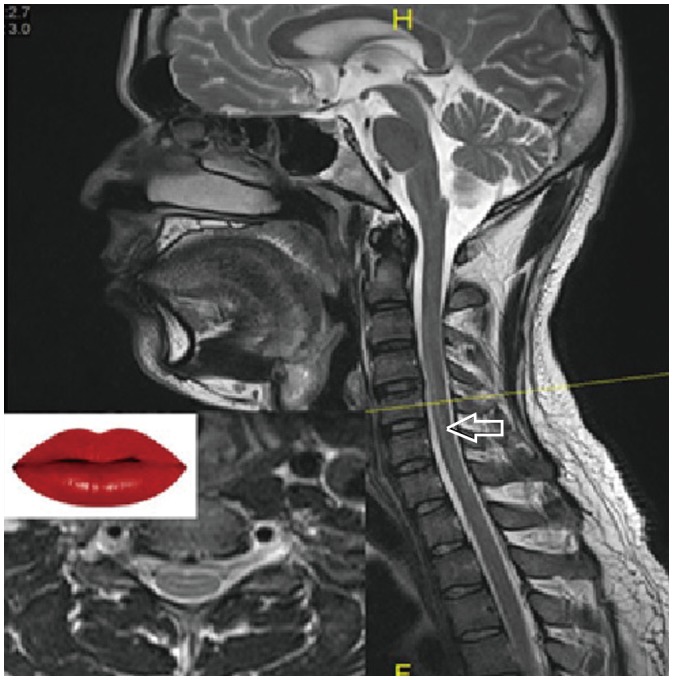
Fig. 1 T2-weighted sagittal image of MRI of cervical spine showing apparent signal changes of truncation artifact (shown by arrow) mimicking pathology with corresponding level in axial section (inset) showing positive ‘lip sign.’
Reference
-
1. Krupa K, Bekiesin´ska-Figatowska M. Artifacts in magnetic resonance imaging. Pol J Radiol. 2015; 80:93–106. PMID: 25745524.
Article2. Morelli JN, Runge VM, Ai F, Attenberger U, Vu L, Schmeets SH, et al. An image-based approach to understanding the physics of MR artifacts. Radiographics. 2011; 31:849–866. PMID: 21571661.
Article3. Budrys T, Veikutis V, Lukosevicius S, Gleizniene R, Monastyreckiene E, Kulakiene I. Artifacts in magnetic resonance imaging: how it can really affect diagnostic image quality and confuse clinical diagnosis. J Vibroeng. 2018; 20:1202–1213.
Article
- Full Text Links
-
- Actions
-
Cited
- CITED
-
- Close
- Share
-
- Similar articles
-
- MR Myelography
- Significance of MRI Cord Signal Patterns in Acute Spinal Trauma
- Acute Spinal-Cord Ischemia: Evolution of MRI Findings
- Morphological Analysis of the Cervical Spinal Cord, Dural Tube, and Spinal Canal by Magnetic Resonance Imaging in Normal Korean Adults
- Spontaneous Herniation of the Thoracic Spinal Cord: A Case Report
