Hepatic Hydatid Disease Causing Gastric Ulcer as a Rare Complication
- Affiliations
-
- 1Department of Gastroenterology, Chonnam National University Medical School, Chonnam National University Hospital, Gwangju, Korea.
- 2Department of Radiology, Chonnam National University Medical School, Chonnam National University Hospital, Gwangju, Korea. kjradsss@gmail.com
- 3Department of Pathology, Chonnam National University Medical School, Chonnam National University Hospital, Gwangju, Korea.
- KMID: 2459026
- DOI: http://doi.org/10.3348/jksr.2019.80.5.987
Abstract
- Hydatid disease in humans is a parasitic disease typically caused by the larvae of Echinococcus granulosus. Although the disease can occur in any body part, it most frequently affects the liver. Hydatid disease is usually diagnosed incidentally and presents with various types of cystic lesion in the infected anatomical locations. Among the many potential complications of hepatic hydatid cysts, including rupture, infection, biliary communication, and peritoneal seeding, spontaneous rupture of the cyst into the hollow viscera is exceptionally rare and has been reported in less than 0.5% of cases. We report the case of a patient with hepatic hydatid disease complicated by spontaneous rupture into stomach causing gastric ulcer and peritoneal seeding.
MeSH Terms
Figure
-
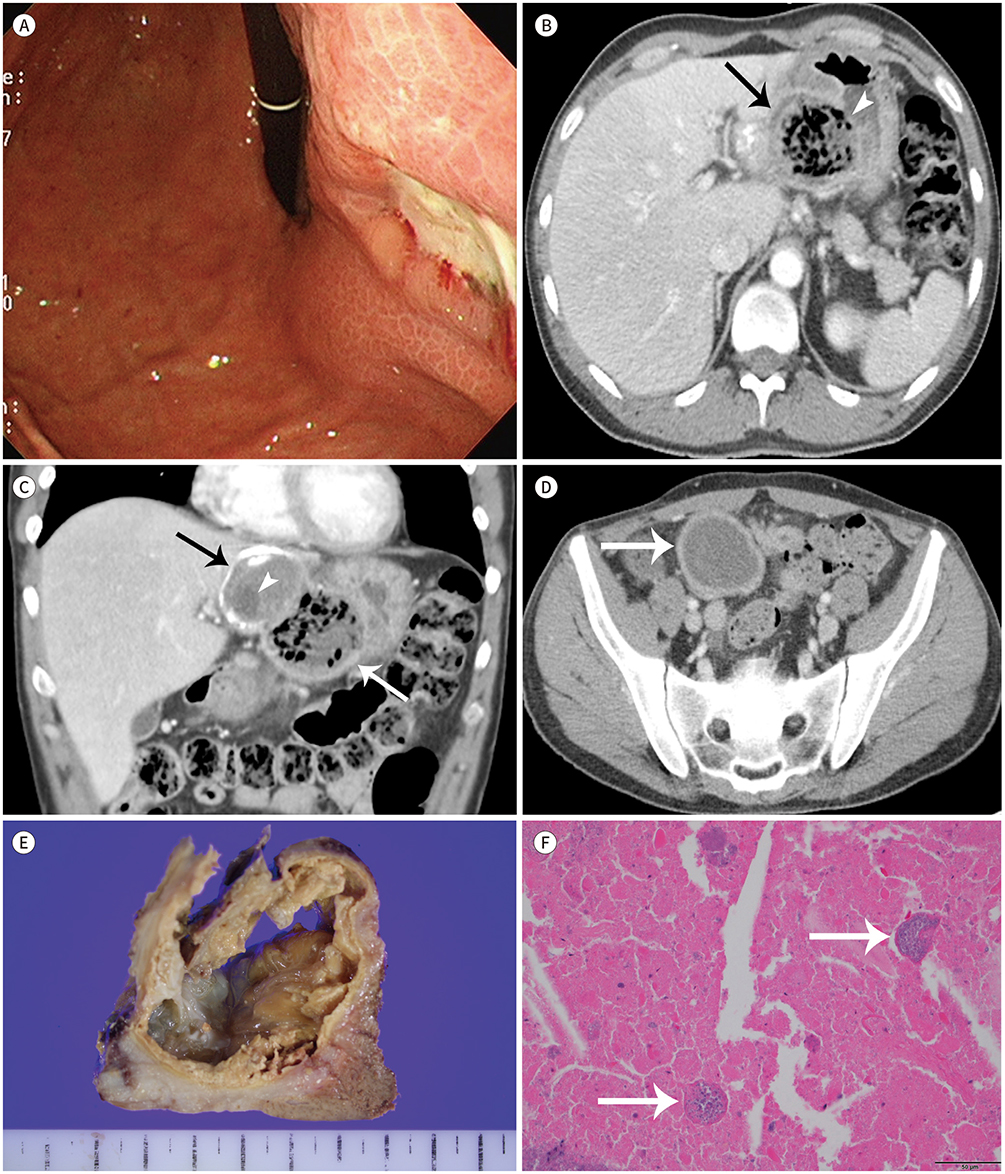
Fig. 1 Hepatic hydatid disease causing gastric ulcer as rare complication in a 49-year-old man. A. An endoscopic image shows a deep ulcer in the lesser curvature side of the gastric upper body surrounded by edematous mucosa. B. An axial contrast-enhanced CT image reveals a round mass (arrow) with mottled air bubbles in the gastrohepatic ligament, causing gastric ulceration (arrowhead). C. A coronal contrast-enhanced CT image demonstrates that the mass (arrows) is located in the hepatic left lateral segment and gastrohepatic ligament, and shows a daughter cyst (arrowhead) and peripheral coarse calcifications. D. An axial contrast-enhanced CT image reveals another cystic mass in the pelvic cavity (arrow) in addition to the hepatic mass. E. Gross surgical specimen demonstrates a turbid fluid-filled cystic mass that contains variable-sized daughter cysts. F. A photomicrograph shows protoscolices (arrows) within necrotic debris (hematoxylin and eosin stain, × 400).
Cited by 1 articles
-
Hepatic Hydatid Cyst: A Case Report
Wan Chul Kim, Jae Uk Shin, Su Sin Jin
Korean J Gastroenterol. 2021;77(1):35-38. doi: 10.4166/kjg.2020.129.
Reference
-
1. Polat P, Kantarci M, Alper F, Suma S, Koruyucu MB, Okur A. Hydatid disease from head to toe. Radiographics. 2003; 23:475–494. quiz 536-537.
Article2. Pedrosa I, Saíz A, Arrazola J, Ferreirós J, Pedrosa CS. Hydatid disease: radiologic and pathologic features and complications. Radiographics. 2000; 20:795–817.
Article3. Zalaquett E, Menias C, Garrido F, Vargas M, Olivares JF, Campos D, et al. Imaging of hydatid disease with a focus on extrahepatic involvement. Radiographics. 2017; 37:901–923.
Article4. Ortega CD, Ogawa NY, Rocha MS, Blasbalg R, Caiado AH, Warmbrand G, et al. Helminthic diseases in the abdomen: an epidemiologic and radiologic overview. Radiographics. 2010; 30:253–267.
Article5. Jain R, Sawhney S, Berry M. Hydatid disease: CT demonstration and follow-up of a cystogastric fistula. AJR Am J Roentgenol. 1992; 158:212.
Article6. Yildiz SY, Berkem H, Hengirmen S. A rare complication of intraabdominal hydatid disease: gastric fistula and recurrent gastric bleeding. Am J Surg. 2010; 200:e59–e60.
Article
- Full Text Links
-
- Actions
-
Cited
- CITED
-
- Close
- Share
-
- Similar articles
-
- Rupture of Right Hepatic Duct into Hydatid Cyst
- Three Cases of Coexistence of Gastric Cancer and Duodenal Ulcer
- An imported case of hepatic unilocular hydatid disease
- Correlation between peptic ulcer and hepatic cirrhosis accompanying ascites
- First Successful Puncture, Aspiration, Injection, and Re-Aspiration of Hydatid Cyst in the Liver Presenting with Anaphylactic Shock in Korea
