Application of the foramina of the trigeminal nerve as landmarks for analysis of craniofacial morphology
- Affiliations
-
- 1Department of Orthodontics, College of Dentistry, Gangneung-Wonju National University, Gangneung, Korea. dschoi@gwnu.ac.kr
- KMID: 2458621
- DOI: http://doi.org/10.4041/kjod.2019.49.5.326
Abstract
OBJECTIVE
The objective of this study was to develop new parameters based on the foramina of the trigeminal nerve and to compare them with the conventional cephalometric parameters in different facial skeletal types.
METHODS
Cone-beam computed tomography (CBCT) scans and cephalograms from 147 adult patients (57 males and 90 females; mean age, 26.1 years) were categorized as Class I (1°< ANB < 3°), Class II (ANB > 5°), and Class III (ANB <−1°). Seven foramina in the craniofacial area"”foramen rotundum (Rot), foramen ovale (Ov), infraorbital foramen, greater palatine foramen, incisive foramen (IF), mandibular foramen (MDF), and mental foramen (MTF)"”were identified in the CBCT images. Various linear, angular, and ratio parameters were compared between the groups by using the foramina, and the relationship between the new parameters and the conventional cephalometric parameters was assessed.
RESULTS
The distances between the foramina in the cranial base did not differ among the three groups. However, the Rot-IF length was shorter in female Class III patients, while the Ov-MTF length, MDF-MTF length, and Ov-MDF length were shorter in Class II patients than in Class III patients of both sexes. The MDF-MTF/FH plane angle was larger in Class II patients than in Class III patients of both sexes. Most parameters showed moderate to high correlations, but the Ov-MDF-MTF angle showed a relatively low correlation with the gonial angle.
CONCLUSIONS
The foramina of the trigeminal nerve can be used to supplement assessments based on the conventional skeletal landmarks on CBCT images.
Keyword
MeSH Terms
Figure
-
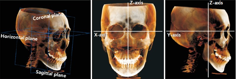
Figure 1 Three-dimensional reconstruction and orientation of the cone-beam computed tomography image with three reference planes and three axes. The origin was set at the sella.
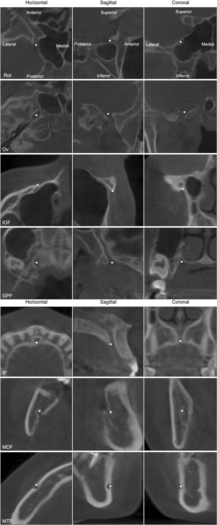
Figure 2 Description of landmarks in cone-beam computed tomography images. The images in each column have the same orientation as the corresponding images in the first row.Rot, Foramen rotundum; Ov, foramen ovale; IOF, infraorbital foramen; GPF, greater palatine foramen; IF, incisive foramen; MDF, mandibular foramen; MTF, mental foramen.
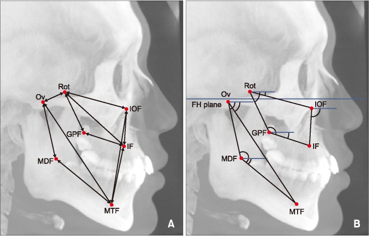
Figure 3 Linear (A) and angular (B) parameters in cone-beam computed tomography (CBCT) images. Identification of the landmark and measurements were performed three-dimensionally. To visualize the location of the landmarks and measurement parameters, they were simply drawn in the lateral maximum projection image of CBCT.FH, Frankfort horizontal plane. See Table 2 for definition of each landmark.
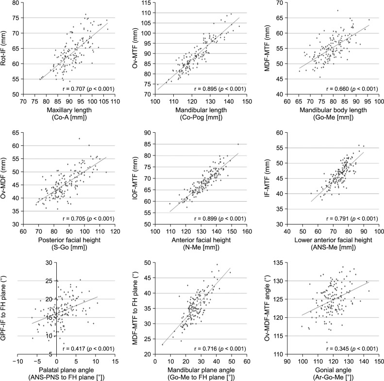
Figure 4 Spearman correlation analysis between conventional cephalometric parameters and the new cone-beam computed tomography parameters using foramina in maxillomandibular analysis.See Tables 2 and 3 for definitions of each landmark or measurement.
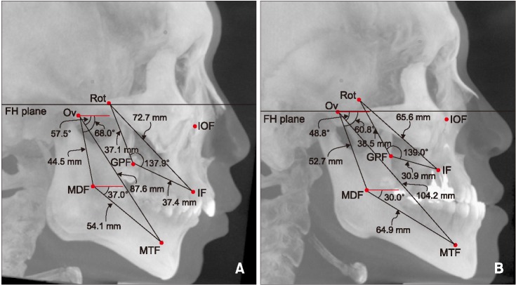
Figure 5 The locations of landmarks and measurements were visualized in the lateral maximum projection image of cone-beam computed tomography. Values of linear and angular parameters as measured in a 22-year-old male patient with skeletal Class II malocclusion (A), and a 23-year-old male patient with skeletal Class III malocclusion (B).FH, Frankfort horizontal plane. See Table 2 for definition of each landmark.
Reference
-
1. Downs WB. The role of cephalometrics in orthodontic case analysis and diagnosis. Am J Orthod. 1952; 38:162–182.
Article2. Ricketts RM. Cephalometric synthesis: an exercise in stating objectives and planning treatment with tracings of the head roentgenogram. Am J Orthod. 1960; 46:647–673.3. Ricketts RM. A foundation for cephalometric communication. Am J Orthod. 1960; 46:330–357.
Article4. Steiner CC. Cephalometrics for you and me. Am J Orthod. 1953; 39:729–755.
Article5. Steiner CC. Cephalometrics in clinical practice. Angle Orthod. 1959; 29:8–29.6. Adams JW. Correction of error in cephalometric roentgenograms. Angle Orthod. 1940; 10:3–13.7. Graber TM. Problems and limitations of cephalometric analysis in orthodontics. J Am Dent Assoc. 1956; 53:439–454. PMID: 13366566.
Article8. Houston WJ. The analysis of errors in orthodontic measurements. Am J Orthod. 1983; 83:382–390. PMID: 6573846.
Article9. Captier G, Lethuilier J, Oussaid M, Canovas F, Bonnel F. Neural symmetry and functional asymmetry of the mandible. Surg Radiol Anat. 2006; 28:379–386. PMID: 16568217.
Article10. Kim SJ, Lee KJ, Lee SH, Baik HS. Morphologic relationship between the cranial base and the mandible in patients with facial asymmetry and mandibular prognathism. Am J Orthod Dentofacial Orthop. 2013; 144:330–340. PMID: 23992805.
Article11. Lagravère MO, Gordon JM, Flores-Mir C, Carey J, Heo G, Major PW. Cranial base foramen location accuracy and reliability in cone-beam computerized tomography. Am J Orthod Dentofacial Orthop. 2011; 139:e203–e210. PMID: 21392663.
Article12. Ludlow JB, Laster WS, See M, Bailey LJ, Hershey HG. Accuracy of measurements of mandibular anatomy in cone beam computed tomography images. Oral Surg Oral Med Oral Pathol Oral Radiol Endod. 2007; 103:534–542. PMID: 17395068.
Article13. Cutright B, Quillopa N, Schubert W. An anthropometric analysis of the key foramina for maxillofacial surgery. J Oral Maxillofac Surg. 2003; 61:354–357. PMID: 12618976.
Article14. Scammon RE. The measurement of the body in childhood. In : Harris J, Jackson C, Paterson D, Scammon RE, editors. The measurement of man. Minneapolis: University of Minnesota;1930.15. Björk A. Prediction of mandibular growth rotation. Am J Orthod. 1969; 55:585–599. PMID: 5253957.
Article16. Gudmundsson K, Rhoton AL Jr, Rushton JG. Detailed anatomy of the intracranial portion of the trigeminal nerve. J Neurosurg. 1971; 35:592–600. PMID: 5120007.
Article17. Gray H, Warwick R, Williams PL. Gray's anatomy. 35th ed. London: Longman;1973.18. Dahlberg G. Statistical methods for medical and biological students. New York: Interscience Publications;1940.19. Behrents RG, Johnston LE Jr. The influence of the trigeminal nerve on facial growth and development. Am J Orthod. 1984; 85:199–206. PMID: 6584031.
Article20. Gardner DE, Luschei ES, Joondeph DR. Alterations in the facial skeleton of the guinea pig following a lesion of the trigeminal motor nucleus. Am J Orthod. 1980; 78:66–80. PMID: 6930172.
Article21. Moss ML. Neurotrophic processes in orofacial growth. J Dent Res. 1971; 50:1492–1494. PMID: 5289054.
Article22. Moss ML. An introduction to the neurobiology of oro-facial growth. Acta Biotheor. 1972; 21:236–259. PMID: 4119332.
Article23. Pimenidis MZ, Gianelly AA. Class III malocclusion produced by oral facial sensory deprivation in the rat. Am J Orthod. 1977; 71:94–102. PMID: 264366.
Article24. Sarnat BG, Feigenbaum JA, Krogman WM. Adult monkey coronoid process after resection of trigeminal nerve motor root. Am J Anat. 1977; 150:129–137. PMID: 412408.
Article25. Kasai K, Moro T, Kanazawa E, Iwasawa T. Relationship between cranial base and maxillofacial morphology. Eur J Orthod. 1995; 17:403–410. PMID: 8529753.
Article26. Sassouni V. A classification of skeletal facial types. Am J Orthod. 1969; 55:109–123. PMID: 5249177.
Article27. Schudy FF. Vertical growth versus anteroposterior growth as related to function and treatment. Angle Orthod. 1964; 34:75–93.28. Sanborn RT. Differences between the facial skeletal patterns of Class III malocclusion and normal occlusion. Angle Orthod. 1955; 25:208–222.
- Full Text Links
-
- Actions
-
Cited
- CITED
-
- Close
- Share
-
- Similar articles
-
- THE DIAGNOSIS OF TRIGEMINAL NERVE INJURY IN FACIAL TRAUMA BY TRIGEMINAL SOMATOSENSORY EVOKED POTENTIAL
- Distribution of the lingual foramina in mandibular cortical bone in Koreans
- Isolated Trigeminal Sensory Change due to Lateral Pontine Hemorrhage Extending to the Trigeminal Nerve Root
- A Case of Secondary Trigeminal Neuralgia Caused by Infected Postoperative Maxillary Cyst
- Role of neuron and non-neuronal cell communication in persistent orofacial pain
