Evaluation of skeletal maturity in the cervical vertebrae and hand-wrist in relation to vertical facial types
- Affiliations
-
- 1Department of Orthodontics, Yonsei University College of Dentistry, Seoul, Korea. hwang@yuhs.ac
- 2Institute of Craniofacial Deformity, Yonsei University College of Dentistry, Seoul, Korea.
- 3Department of Orthodontics, Gangnam Severance Dental Hospital, Yonsei University College of Dentistry, Seoul, Korea.
- KMID: 2458620
- DOI: http://doi.org/10.4041/kjod.2019.49.5.319
Abstract
OBJECTIVE
To evaluate differences in skeletal maturity in relation to vertical facial types and to compare differences in the skeletal maturity of the cervical vertebrae and hand-wrist in females.
METHODS
This study included 59 females aged 7 to 9 years with skeletal Class I malocclusion. The participants were categorized into three groups (low, normal, and high) according to the mandibular plane angle. Skeletal maturity was measured using skeletal maturity indicators (SMIs) and the Tanner-Whitehouse III (TW3) method on hand-wrist radiographs and by using cervical vertebrae maturation indicators (CVMIs) on lateral cephalometric radiographs.
RESULTS
The SMI was higher in the high-angle group than in the low-angle group (p = 0.014). The median TW3 bone age was 11.4 months higher in the high-angle group than in the low-angle group (p = 0.032). There was no significant difference in CVMI among the three groups. Skeletal maturity showed a weakly positive correlation with the mandibular plane angle (SMI, r = 0.391; TW3, r = 0.333; CVMI, r = 0.259).
CONCLUSIONS
The skeletal maturity of the hand-wrist in females with a high mandibular plane angle was higher than that in females with a low mandibular plane angle. Obtaining additional hand-wrist radiographs may facilitate evaluation of skeletal maturity of females. In females with a high mandibular angle, the time to commence orthodontic treatment may be earlier than that in females with a low mandibular angle.
Keyword
Figure
-
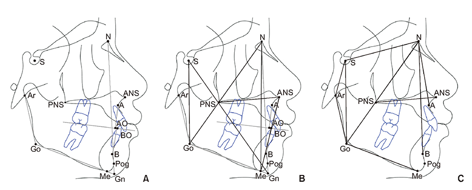
Figure 1 Cephalometric analysis. A, Cephalometric landmarks. S, Sella turcica; N, nasion; Ar, articulare; ANS, anterior nasal spine; PNS, posterior nasal spine; A, point A; B, point B; AO, the point perpendicular to the occlusal plane at point A; BO, the point perpendicular to the occlusal plane at point B; Go, constructed gonion; Me, menton; Pog, pogonion; Gn, mechanical gnathion. B, Linear measurements. AO-BO, Wits; N-Me, anterior facial height; ANS-Me, lower anterior facial height; S-Go, posterior facial height; N-Go, facial depth; S-Gn, facial length. C, Angular measurements. SNA, The relationship of the maxilla to the cranial base; SNB, the relationship of the mandible to the cranial base; ANB, the relationship between the maxilla and the mandible; SN-GoMe, mandibular plane angle; Ar-Go-Me, gonial angle; Ar-Go-N, upper half of the gonial angle; N-Go-Me, lower half of the gonial angle.
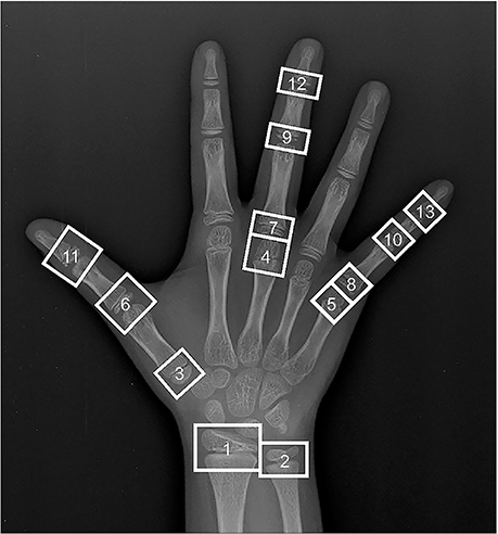
Figure 2 Thirteen measurement points on the hand using the Tanner–White III method. 1, Radius; 2, ulna; 3, metacarpal I; 4, metacarpal III; 5, metacarpal V; 6, proximal phalange I; 7, proximal phalange III; 8, proximal phalange V; 9, middle phalange III; 10, middle phalange V; 11, distal phalange I; 12, distal phalange III; 13, distal phalange V.
Cited by 1 articles
-
Evaluation of cephalometric characteristics and skeletal maturation of the cervical vertebrae and hand-wrist in girls with central precocious puberty
Sung-Tae Kang, Sung-Hwan Choi, Kyung-Ho Kim, Chung-Ju Hwang
Korean J Orthod. 2020;50(3):181-187. doi: 10.4041/kjod.2020.50.3.181.
Reference
-
1. Fishman LS. Chronological versus skeletal age, an evaluation of craniofacial growth. Angle Orthod. 1979; 49:181–189.2. Demirjian A, Buschang PH, Tanguay R, Patterson DK. Interrelationships among measures of somatic, skeletal, dental, and sexual maturity. Am J Orthod. 1985; 88:433–438.
Article3. Flores-Mir C, Nebbe B, Major PW. Use of skeletal maturation based on hand-wrist radiographic analysis as a predictor of facial growth: a systematic review. Angle Orthod. 2004; 74:118–124.4. Fishman LS. Radiographic evaluation of skeletal maturation. A clinically oriented method based on hand-wrist films. Angle Orthod. 1982; 52:88–112.5. Tanner JM, Healy MJR, Goldstein H, Cameron N. Assessment of skeletal maturity and prediction of adult height (TW3 method). Philadelphia: Saunders;2001.6. Hägg U, Taranger J. Skeletal stages of the hand and wrist as indicators of the pubertal growth spurt. Acta Odontol Scand. 1980; 38:187–200.
Article7. Hassel B, Farman AG. Skeletal maturation evaluation using cervical vertebrae. Am J Orthod Dentofacial Orthop. 1995; 107:58–66.
Article8. Cericato GO, Bittencourt MA, Paranhos LR. Validity of the assessment method of skeletal maturation by cervical vertebrae: a systematic review and meta-analysis. Dentomaxillofac Radiol. 2015; 44:20140270.
Article9. Nanda SK. Patterns of vertical growth in the face. Am J Orthod Dentofacial Orthop. 1988; 93:103–116.
Article10. Verulkar A, Singla P, Patil HA, Tekale PD. Assessment of skeletal and dental maturity indicators and comparison of maturity indicators in vertical and horizontal growth pattern individuals with normal growth pattern individuals. Int J Orthod Rehabil. 2017; 8:108–111.
Article11. Park IO, Sohn BH. A longitudinal study of the changes in the anteroposterior apical base relationship between 6 and 13 years of age. Korean J Orthod. 1989; 19:137–151.12. Kim KH, Choy KC, Lee JY. A study on horizontal reference planes in lateral cephalogram in Korean children. Korean J Orthod. 1999; 29:251–264.13. Prion S, Haerling KA. Making sense of methods and measurement: Spearman-rho ranked-order correlation coefficient. Clin Simul Nurs. 2014; 10:535–536.
Article14. Berberoğlu M. Precocious puberty and normal variant puberty: definition, etiology, diagnosis and current management. J Clin Res Pediatr Endocrinol. 2009; 1:164–174.
Article15. Lee HK, Choi SH, Fan D, Jang KM, Kim MS, Hwang CJ. Evaluation of characteristics of the craniofacial complex and dental maturity in girls with central precocious puberty. Angle Orthod. 2018; 88:582–589.
Article16. Kim KH. A longitudinal study on the skeletal maturity of the hand and wrist among various malocclusion groups (I). Korean J Orthod. 1999; 29:183–195.17. Kim KH, Baik HS, Son ES. A study on menarche and skeletal maturity among various malocclusion groups. Korean J Orthod. 1998; 28:581–589.18. Neves LS, Pinzan A, Janson G, Canuto CE, de Freitas MR, Cançado RH. Comparative study of the maturation of permanent teeth in subjects with vertical and horizontal growth patterns. Am J Orthod Dentofacial Orthop. 2005; 128:619–623.
Article19. Ricketts RM. Cephalometric analysis and synthesis. Angle Orthod. 1961; 31:141–156.20. Ashizawa K, Kumakura C, Zhou X, Jin F, Cao J. RUS skeletal maturity of children in Beijing. Ann Hum Biol. 2005; 32:316–325.
Article21. Kannus P, Haapasalo H, Sievänen H, Oja P, Vuori I. The site-specific effects of long-term unilateral activity on bone mineral density and content. Bone. 1994; 15:279–284.
Article22. Martin DD, Neuhof J, Jenni OG, Ranke MB, Thodberg HH. Automatic determination of left- and right-hand bone age in the First Zurich Longitudinal Study. Horm Res Paediatr. 2010; 74:50–55.
Article23. Shin YH, Chyang I, Kang HY. A comparison of bone maturation between dominant hand and non-dominant hand of boys in childhood by means of Tanner-Whitehouse 3 (TW3) method. Korean J Sports Sci. 2014; 23:1459–1466.
- Full Text Links
-
- Actions
-
Cited
- CITED
-
- Close
- Share
-
- Similar articles
-
- Correlations between dental and skeletal maturity
- A study on the degrees of skeletal maturity of cervical vertebrae and hand-and-wrist in skeletal Class III malocclusions
- Evaluation of the skeletal maturity using the cervical vertebrae and hand-wrist rakiographs
- Evaluation of Skeletal and Dental Maturity in Relation to Vertical Facial Types and the Sex of Growing Children
- Skeletal maturation evaluation using mandibular third molar development in adolescents
