The Predictable Factors of the Postoperative Kyphotic Change of Sagittal Alignment of the Cervical Spine after the Laminoplasty
- Affiliations
-
- 1Department of Neurosurgery, Pusan National University Yangsan Hospital, Yangsan, Korea. md6576@naver.com
- 2Research Institute for Convergence of Biomedical Science and Technology, Pusan National University Yangsan Hospital, Yangsan, Korea.
- 3Department of Neurosurgery, Pusan National University School of Medicine, Yangsan, Korea.
- KMID: 2457965
- DOI: http://doi.org/10.3340/jkns.2017.0505.007
Abstract
OBJECTIVE
Laminoplasty is an effective surgical method for treating cervical degenerative disease. However, postoperative complications such as kyphosis, restriction of neck motion, and instability are often reported. Despite sufficient preoperative lordosis, this procedure often aggravates the lordotic curve of the cervical spine and straightens cervical alignment. Hence, it is important to examine preoperative risk factors associated with postoperative kyphotic alignment changes. Our study aimed to investigate preoperative radiologic parameters associated with kyphotic deformity post laminoplasty.
METHODS
We retrospectively reviewed the medical records of 49 patients who underwent open door laminoplasty for cervical spondylotic myelopathy (CSM) or ossification of the posterior longitudinal ligament (OPLL) at Pusan National University Yangsan Hospital between January 2011 and December 2015. Inclusion criteria were as follows: 1) preoperative diagnosis of OPLL or CSM, 2) no previous history of cervical spinal surgery, cervical trauma, tumor, or infection, 3) minimum of one-year follow-up post laminoplasty with proper radiologic examinations performed in outpatient clinics, and 4) cases showing C7 and T1 vertebral body in the preoperative cervical sagittal plane. The radiologic parameters examined included C2-C7 Cobb angles, T1 slope, C2-C7 sagittal vertical axis (SVA), range of motion (ROM) from C2-C7, segmental instability, and T2 signal change observed on magnetic resonance imaging (MRI). Clinical factors examined included preoperative modified Japanese Orthopedic Association scores, disease classification, duration of symptoms, and the range of operation levels.
RESULTS
Mean preoperative sagittal alignment was 13.01° lordotic; 6.94° lordotic postoperatively. Percentage of postoperative kyphosis was 80%. Patients were subdivided into two groups according to postoperative Cobb angle change; a control group (n=22) and kyphotic group (n=27). The kyphotic group consisted of patients with more than 5° kyphotic angle change postoperatively. There were no differences in age, sex, C2-C7 Cobb angle, T1 slope, C2-C7 SVA, ROM from C2-C7, segmental instability, or T2 signal change. Multiple regression analysis revealed T1 slope had a strong relationship with postoperative cervical kyphosis. Likewise, correlation analysis revealed there was a statistical significance between T1 slope and postoperative Cobb angle change (p=0.035), and that there was a statistically significant relationship between T1 slope and C2-C7 SVA (p=0.001). Patients with higher preoperative T1 slope demonstrated loss of lordotic curvature postoperatively.
CONCLUSION
Laminoplasty has a high probability of aggravating sagittal balance of the cervical spine. T1 slope is a good predictor of postoperative kyphotic changes of the cervical spine. Similarly, T1 slope is strongly correlated with C2-C7 SVA.
Keyword
MeSH Terms
-
Ambulatory Care Facilities
Animals
Asian Continental Ancestry Group
Busan
Cervical Vertebrae
Classification
Congenital Abnormalities
Diagnosis
Female
Follow-Up Studies
Gyeongsangnam-do
Humans
Kyphosis
Laminoplasty*
Longitudinal Ligaments
Lordosis
Magnetic Resonance Imaging
Medical Records
Methods
Neck
Orthopedics
Postoperative Complications
Range of Motion, Articular
Retrospective Studies
Risk Factors
Spinal Cord Diseases
Spine*
Spondylosis
Figure
-

Fig. 1 Measurement of radiologic parameters, including C2–C7 SVA, C2–C7 lordosis and T1 slope in cervical lateral radiographs. SVA: sagittal vertical axis.

Fig. 2 Scatter plots showing a positive correlation between T1 slope and loss of cervical lordosis (r=0.302). CA: cobb angle.
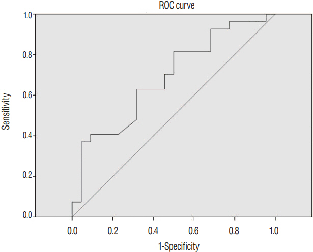
Fig. 3 ROC analysis was performed to determine the cut-off value of T1 slope that induced significant postoperative kyphotic change. T1 slope above 29° could serve as a threshold for significantly increased risk of postoperative kyphotic change (p=0.026, area under curve=0.687, sensitivity=63%, specificity=69%). ROC: receiver operating characteristic.
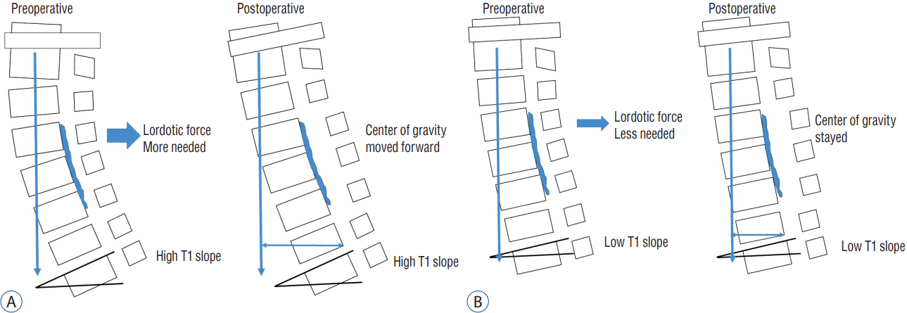
Fig. 4 Schematic image of alteration of cervical lordosis after laminoplasty in patients with OPLL. A: Patients with high T1 slope had needed more lordotic force. Postoperatively, center of gravity moved forward and cervical spine accelerated kyphotic. B: Patients with low T1 slope had relatively less influence of the operation than high T1 slope. OPLL: ossification of the posterior longitudinal ligament.
Cited by 1 articles
-
Preoperative Radiological Parameters to Predict Clinical and Radiological Outcomes after Laminoplasty
Su Hun Lee, Dong Wuk Son, Jun Jae Shin, Yoon Ha, Geun Sung Song, Jun Seok Lee, Sang Weon Lee
J Korean Neurosurg Soc. 2021;64(5):677-692. doi: 10.3340/jkns.2020.0294.
Reference
-
References
1. Cho JH, Ha JK, Kim DG, Song KY, Kim YT, Hwang CJ, et al. Does preoperative T1 slope affect radiological and functional outcomes after cervical laminoplasty? Spine (Phila Pa 1976). 39:E1575–E1581. 2014.
Article2. Dugoni DE, Mancarella C, Landi A, Tarantino R, Ruggeri AG, Delfini R. Post laminoplasty cervical kyphosis-case report. Int J Surg Case Rep. 5:853–857. 2014.
Article3. Ellwitz J, Roberto R, Gupta M, Mohan V, Klineberg E. Patient and surgeon factors associated with postoperative kyphosis after laminoplasty. Evid Based Spine Care J. 2:53–54. 2011.
Article4. Kang SH, Rhim SC, Roh SW, Jeon SR, Baek HC. Postlaminoplasty cervical range of motion: early results. J Neurosurg Spine. 6:386–390. 2007.
Article5. Kim B, Yoon DH, Ha Y, Yi S, Shin DA, Lee CK, et al. Relationship between T1 slope and loss of lordosis after laminoplasty in patients with cervical ossification of the posterior longitudinal ligament. Spine J. 16:219–225. 2016.
Article6. Kim TH, Lee SY, Kim YC, Park MS, Kim SW. T1 slope as a predictor of kyphotic alignment change after laminoplasty in patients with cervical myelopathy. Spine (Phila Pa 1976). 38:E992–E997. 2013.
Article7. Knott PT, Mardjetko SM, Techy F. The use of the T1 sagittal angle in predicting overall sagittal balance of the spine. Spine J. 10:994–998. 2010.
Article8. Matsumoto M, Chiba K, Toyama Y. Surgical treatment of ossification of the posterior longitudinal ligament and its outcomes: posterior surgery by laminoplasty. Spine (Phila Pa 1976). 37:E303–E308. 2012.9. Michael KW, Neustein TM, Rhee JM. Where should a laminoplasty start? The effect of the proximal level on post-laminoplasty loss of lordosis. Spine J. 16:737–741. 2016.
Article10. Sakai K, Yoshii T, Hirai T, Arai Y, Shinomiya K, Okawa A. Impact of the surgical treatment for degenerative cervical myelopathy on the preoperative cervical sagittal balance: a review of prospective comparative cohort between anterior decompression with fusion and laminoplasty. Eur Spine J. 26:104–112. 2017.
Article11. Suk KS, Kim KT, Lee JH, Lee SH, Lim YJ, Kim JS. Sagittal alignment of the cervical spine after the laminoplasty. Spine (Phila Pa 1976). 32:E656–E660. 2007.
Article12. Tang JA, Scheer JK, Smith JS, Deviren V, Bess S, Hart RA, et al. The impact of standing regional cervical sagittal alignment on outcomes in posterior cervical fusion surgery. Neurosurgery. 71:662–669. discussion 669. 2012.
Article13. Uchida K, Nakajima H, Sato R, Yayama T, Mwaka ES, Kobayashi S, et al. Cervical spondylotic myelopathy associated with kyphosis or sagittal sigmoid alignment: outcome after anterior or posterior decompression. J Neurosurg Spine. 11:521–528. 2009.
Article14. White AA 3rd, Panjabi MM. Update on the evaluation of instability of the lower cervical spine. Instr Course Lect. 36:513–520. 1987.15. Yang BS, Lee SK, Song KS, Yoon SP, Jang G, Lee CC, et al. The use of T1 sagittal angle in predicting cervical disc degeneration. Asian Spine J. 9:757–761. 2015.
Article
- Full Text Links
-
- Actions
-
Cited
- CITED
-
- Close
- Share
-
- Similar articles
-
- Changes of Range of Motion and Sagittal Alignment of the Cervical Spine after Laminoplasty
- Cervical Sagittal Alignment: Literature Review and Future Directions
- Kyphotic Neck and Correlation With Clinical Outcomes
- The Sagittal Balance of Cervical Spine : Comprehensive Review of Recent Update
- Surgical and Functional Outcomes of Expansive Open-Door Laminoplasty for Patients With Mild Kyphotic Cervical Alignment
