Desmoid-Type Fibromatosis Associated with Silicone Breast Implants
- Affiliations
-
- 1Department of Radiology, Yeungnam University College of Medicine, Daegu, Korea. air1986@naver.com
- 2Department of Pathology, Yeungnam University College of Medicine, Daegu, Korea.
- KMID: 2457462
- DOI: http://doi.org/10.3348/jksr.2019.80.4.804
Abstract
- Desmoid-type fibromatosis (extra-abdominal desmoid tumor) arising in the breast is a rare benign disease entity, accounting for 0.2% of all breast tumors. The etiology of mammary fibromatosis is unknown, but has been reported to be associated with genetic factors, hormonal effects, Gardner syndrome, breast trauma or surgery, and breast implants. Fibromatosis associated with breast implants is extremely rare, and only 34 cases have been reported so far. We report the ultrasonographic, computed tomographic, and pathologic findings of breast fibromatosis in a 29-year-old woman, who underwent bilateral augmentation mammoplasty with silicone implants.
MeSH Terms
Figure
-
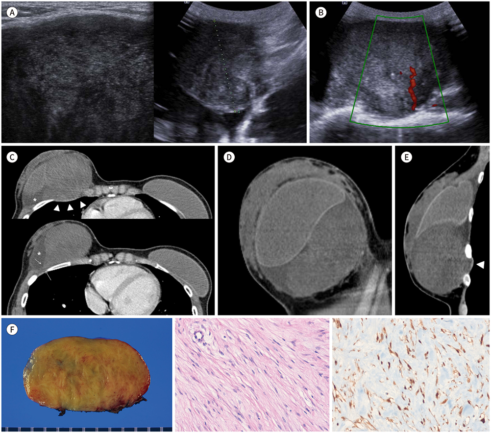
Fig. 1 Radiologic and pathologic findings of desmoid-type fibromatosis associated with silicone breast implants in a 29-year-old woman. A. Ultrasonography images show an oval-shaped, circumscribed marginated heterogeneous hypoechoic mass beneath the pectoralis major muscle of the right breast. The lesion is classified as BI-RADS category 4A based on ultrasound image findings. B. Color Doppler study shows internal vascularity within the mass. C–E. Axial (C), sagittal (D), and coronal (E) images obtained with contrast-enhanced CT of the chest reveal a well-defined oval-shaped mass, measuring 10.5 × 6.5 × 6.0 cm, that is in direct contact with the inferomedial aspect of the right breast implant. The mass extends to the thorax with chest wall invasion and indentation of the adjacent pleura (arrowheads). The mass shows spindle-shaped margins with a characteristic linear tail (arrows) extending along the fascial plane of the chest wall. Small amounts of fluid collection (*) can be observed in the capsule between the mass and implant. F. Gross specimen (left panel) demonstrates a well-defined yellowish- to gray-colored mass measuring 10.0 × 6.5 × 6.0 cm. Microscopic findings (middle panel) demonstrate spindle-shaped tumor cells arranged in a long fascicular pattern (hematoxylin and eosin stain, × 200). The tumor cells show nuclear staining for β-catenin (immunohistochemical stain × 200) (right panel). BI-RADS = Breast Imaging Reporting and Data System
Reference
-
1. Neuman HB, Brogi E, Ebrahim A, Brennan MF, Van Zee KJ. Desmoid tumors (fibromatoses) of the breast: a 25-year experience. Ann Surg Oncol. 2008; 15:274–280.
Article2. Glazebrook KN, Reynolds CA. Mammary fibromatosis. AJR Am J Roentgenol. 2009; 193:856–860.
Article3. Seo YN, Park YM, Yoon HK, Lee SJ, Choo HJ, Ryu JH. Breast fibromatosis associated with breast implants. Jpn J Radiol. 2015; 33:591–597.
Article4. Alanis L, Roth R, Lerman N, Barroeta JE, Germaine P. Radiologic images of an aggressive implant-associated fibromatosis of the breast and chest wall: case report and review of the literature. Radiol Case Rep. 2017; 12:431–438.
Article5. Shim HS, Kim SJ, Kim OH, Jung HK, Kim SJ, Kim W, et al. Fibromatosis associated with silicone breast implant: ultrasonography and MR imaging findings. Breast J. 2014; 20:645–649.
Article6. Murphey MD, Ruble CM, Tyszko SM, Zbojniewicz AM, Potter BK, Miettinen M. From the archives of the AFIP: musculoskeletal fibromatoses: radiologic-pathologic correlation. Radiographics. 2009; 29:2143–2173.7. Kim HJ, Lee KH, Kim JH, Shim MK, Lee JS, Choi C. Fibromatosis of the breast: a case report. Korean J Pathol. 2005; 39:137–139.
- Full Text Links
-
- Actions
-
Cited
- CITED
-
- Close
- Share
-
- Similar articles
-
- Recurring Fibromatosis of Breast Following Tumorectomy: A Case Report
- Desmoid Tumor of the Facet Joint: A Case Report
- Reoperative Augmentation Mammoplasty of Saline Implants with Cohesive Silicone Gel Implants: A Personal Review
- Desmoid Type Fibromatosis in the Facet Joint of Lumbar Spine: Case Report and Review of Literature
- A Retrospective Analysis of Ruptured Breast Implants
