Functional impression technique using temporary denture for rehabilitation of severely atrophic maxillary and mandibular ridges
- Affiliations
-
- 1Department of Prosthodontics, Gil Medical Center, Gachon University, Incheon, Republic of Korea. bb1018@hanmail.net
- KMID: 2453778
- DOI: http://doi.org/10.4047/jkap.2019.57.3.238
Abstract
- Soft liner is used to functional impression technique when dental stone is immediately poured after taking impression because of viscoelasticity. In this case, a 78-year-old male visited for new dentures. Due to severe resorption of mandibular edentulous ridge, functional impression taking by closed mouth technique was planned. First of all, making maxillary and mandibular provisional dentures was done, and lined by soft liner to rehabilitate pressured maxillary and mandibular edentulous ridge. After this, Functional impression was taken by closed mouth technique using provisional dentures which are transformed to healed maxillary and mandibular edentulous ridge, and final denture were fabricated using maxillary provisional denture as a reference of artificial teeth arrangement. Consequently, restoring a complete edentulous patient with taking functional impression using provisional dentures resulted in recovering satisfying retention and function.
Figure
-

Fig. 1 Initial photographs. (A) Occlusal view of maxilla, (B) Right lateral view, (C) Frontal view, (D) Left lateral view, (E) Occlusal view of mandible, (F) Panoramic radiograph.
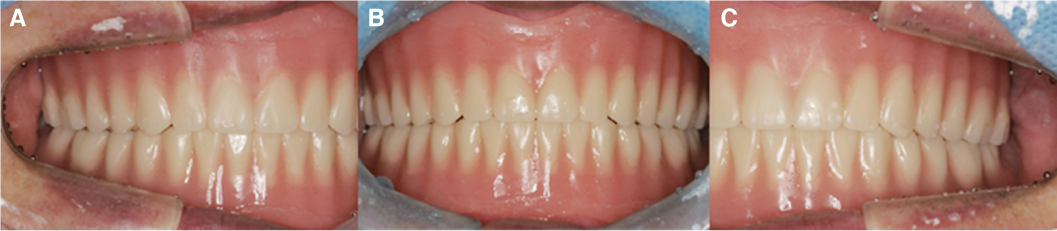
Fig. 2 Provisional denture intraoral view in occlusion. (A) Right lateral view, (B) Frontal view, (C) Left lateral view.
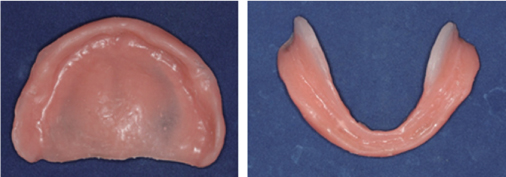
Fig. 3 Final impression taking using Soft liner.

Fig. 4 Facebow transfer using upper provisional denture.
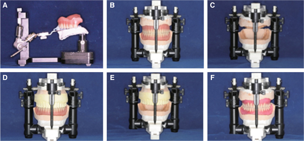
Fig. 5 (A) Facebow transfer using upper provisional denture, (B) Mounting using provisional denture, (C) Jaw relation, (D) Replicate upper provisional denture and mounting with lower provisional denture, (E) Upper provisional denture mounting with mandible, (F) Denture tooth set up.
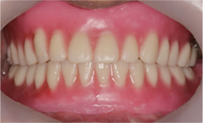
Fig. 6 Wax denture try in.
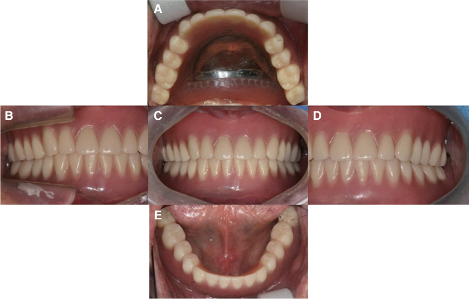
Fig. 7 Intraoral photographs of definitive prosthesis. (A) Upper occlusal view, (B) Right lateral view, (C) Frontal view, (D) Left lateral view, (E) Lower occlusal view.
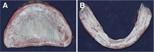
Fig. 8 6 month follow up. (A) Internal fit of upper complete denture, (B) Internal fit of lower complete denture.
Reference
-
1. Chandrasekharan NK, Kunnekel AT, Verma M, Gupta RK. A technique for impressing the severely resorbed mandibular edentulous ridge. J Prosthodont. 2012; 21:215–218.
Article2. Chander S, Hill M, Moore D, Morrow L. Tissue conditioning materials as functional impression materials. Eur J Prosthodont Restor Dent. 2007; 15:67–71.3. Baslas V, Singh SV, Aggarwal H, Kaur S, Singh K, Agarwal KK. A technique for using short term soft liners as complete dentures final impression material. J Oral Biol Craniofac Res. 2014; 4:204–207.
Article4. Wilson HJ. Tissue conditioners and functional impression materials [abridged]. Proc R Soc Med. 1966; 59:1037–1038.
Article5. Gahan MJ, Walmsley AD. The neutral zone impression revisited. Br Dent J. 2005; 198:269–272.
Article6. Kwon KR, Kwon HB, Kim MJ, Kim HJ, Moon HS, Park SW, Park CJ, Song KY, Lee JS, Lee JH, Lee CH, Lim YJ, Chung MK, Chung CH, Jeong CM, Cho IH, Cho HW, Han CH. Prosthodontic treatment for edentulous patients. 2nd ed. Dental Wisdom;2014. p. 139–140.7. Jacobson TE, Krol AJ. A contemporary review of the factors involved in complete dentures. Part II: stability. J Prosthet Dent. 1983; 49:165–172.
Article8. Prithviraj DR, Singh V, Kumar S, Shruti DP. Conservative prosthodontic procedures to improve mandibular denture stability in an atrophic mandibular ridge. J Indian Prosthodont Soc. 2008; 8:178–184.
Article9. Hashem MI. Advances in Soft Denture Liners: An Update. J Contemp Dent Pract. 2015; 16:314–318.
Article10. Kršek H, Dulčić N. Functional impressions in complete denture and overdenture treatment. Acta Stomatol Croat. 2015; 49:45–53.
Article11. Drago CJ. A retrospective comparison of two definitive impression techniques and their associated postinsertion adjustments in complete denture prosthodontics. J Prosthodont. 2003; 12:192–197.
Article12. Wang JC, Hong JM. Tissue conditioner impression technique for edentulous ridges. Chin Dent J. 1998; 17:48–52.13. Chander S, Hill M, Moore D, Morrow L. Tissue conditioning materials as functional impression materials. Eur J Prosthodont Restor Dent. 2007; 15:67–71.14. Hopkins R. The immediate denture. Br Dent J. 1979; 147:71–72.
Article15. McCarthy JA, Moser JB. Tissue conditioning and functional impression materials and techniques. Dent Clin North Am. 1984; 28:239–251.16. Murata H, Hamada T, Djulaeha E, Nikawa H. Rheology of tissue conditioners. J Prosthet Dent. 1998; 79:188–199.
Article17. Murata H, Kawamura M, Hamada T, Saleh S, Kresnoadi U, Toki K. Dimensional stability and weight changes of tissue conditioners. J Oral Rehabil. 2001; 28:918–923.
Article18. Kwon KR, Kwon HB, Kim MJ, Kim HJ, Moon HS, Park SW, Park CJ, Song KY, Lee JS, Lee JH, Lee CH, Lim YJ, Chung MK, Chung CH, Jeong CM, Cho IH, Cho HW, Han CH. Prosthodontic treatment for edentulous patients. 2nd ed. Dental Wisdom;2014. p. 144.19. Basker RM, Davenport JC. Prosthetic treatment of the edentulous patient. 4th ed. Blackwell Munksgaard, a Blackwell Publishing Company;2002. p. 278–279.
- Full Text Links
-
- Actions
-
Cited
- CITED
-
- Close
- Share
-
- Similar articles
-
- Neutral zone approach for rehabilitation of severely atrophic maxillary and mandibular ridges: A case report
- Neutral zone approach and external impression for rehabilitation of severely atrophic maxillary and mandibular ridges: a case report
- Complete denture made with closed-mouth impression technique on severely atrophied edentulous jaw
- Rehabilitation of a patient with atrophic ridges using gothic arch tracing and nonpressure impression: a case report
- Implant-assisted removable partial denture for severely atrophied mandible
