Diagnostic Performance of ¹â¸F-Fluorodeoxyglucose Positron Emission Tomography/CT for Chronic Empyema-Associated Malignancy
- Affiliations
-
- 1Department of Nuclear Medicine, Veterans Health Service Medical Center, Seoul, Korea.
- 2Department of Nuclear Medicine, Samsung Medical Center, Sungkyunkwan University School of Medicine, Seoul, Korea. jynm.choi@samsung.com
- 3Department of Radiology, Samsung Medical Center, Sungkyunkwan University School of Medicine, Seoul, Korea.
- 4Department of Medicine, Samsung Medical Center, Division of Pulmonary and Critical Care Medicine, Sungkyunkwan University School of Medicine, Seoul, Korea.
- 5Department of Thoracic Surgery and Cardiovascular Surgery, Samsung Medical Center, Sungkyunkwan University School of Medicine, Seoul, Korea.
- KMID: 2453079
- DOI: http://doi.org/10.3348/kjr.2018.0843
Abstract
OBJECTIVE
The purpose of this study was to evaluate the diagnostic performance of ¹â¸F-fluorodeoxyglucose positron emission tomography/computed tomography (¹â¸F-FDG PET/CT) for chronic empyema-associated malignancy (CEAM).
MATERIALS AND METHODS
We retrospectively reviewed the ¹â¸F-FDG PET/CT images of 33 patients with chronic empyema, and analyzed the following findings: 1) shape of the empyema cavity, 2) presence of fistula, 3) maximum standardized uptake value (SUV) of the empyema cavity, 4) uptake pattern of the empyema cavity, 5) presence of a protruding soft tissue mass within the empyema cavity, and 6) involvement of adjacent structures. Final diagnosis was determined based on histopathology or clinical follow-up for at least 6 months. The abovementioned findings were compared between the ¹â¸F-FDG PET/CT images of CEAM and chronic empyema. A receiver operating characteristic (ROC) analysis was also performed.
RESULTS
Six lesions were histopathologically proven as malignant; there were three cases of diffuse large B-cell lymphoma, two of squamous cell carcinoma, and one of poorly differentiated carcinoma. Maximum SUV within the empyema cavity (p < 0.001) presence of a protruding soft tissue mass (p = 0.002), and involvement of the adjacent structures (p < 0.001) were significantly different between the CEAM and chronic empyema images. The maximum SUV exhibited the highest diagnostic performance, with the highest specificity (96.3%, 26/27), positive predictive value (85.7%, 6/7), and accuracy (97.0%, 32/33) among all criteria. On ROC analysis, the area under the curve of maximum SUV was 0.994.
CONCLUSION
¹â¸F-FDG PET/CT can be useful for diagnosing CEAM in patients with chronic empyema. The maximum SUV within the empyema cavity is the most accurate ¹â¸F-FDG PET/CT diagnostic criterion for CEAM.
MeSH Terms
Figure
-
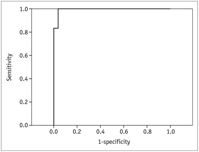
Fig. 1 Receiver operating characteristic curve of maximum standardized uptake value for diagnosing chronic empyema-associated malignancy in patients with chronic empyema.AUC was 0.994 ± 0.010 (p < 0.001). AUC = area under curve
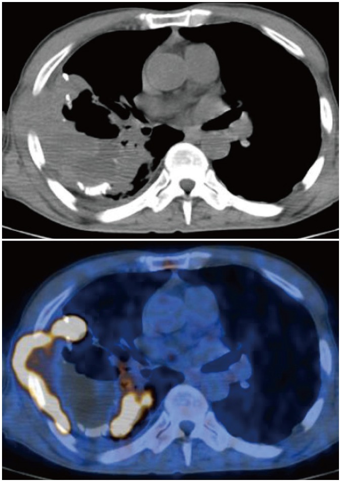
Fig. 2 Non-contrast CT and fused PET/CT images of 68-year-old man with 40-year history of chronic tuberculous empyema.Highly hypermetabolic lesion (maximum SUV = 29.6) involving adjacent chest wall is present within empyema cavity. Lesion was histopathologically proven as diffuse large B-cell lymphoma. CT = computed tomography, PET = positron emission tomography, SUV = standardized uptake value
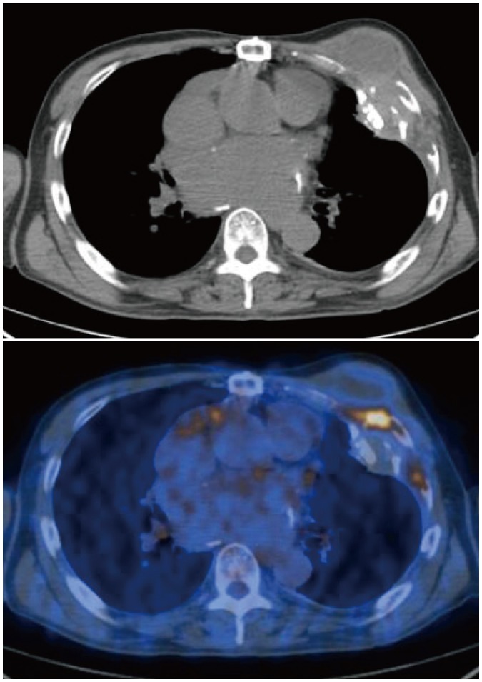
Fig. 3 Non-contrast CT and fused PET/CT images of 69-year-old man with chronic empyema.Moderately hypermetabolic lesion (maximum SUV = 7.0) involving adjacent rib is seen. Lesion was histopathologically proven as chronic active inflammation.
Reference
-
1. Iuchi K, Ichimiya A, Akashi A, Mizuta T, Lee YE, Tada H, et al. Non-Hodgkin's lymphoma of the pleural cavity developing from long-standing pyothorax. Cancer. 1987; 60:1771–1775. PMID: 3308054.
Article2. Iuchi K, Aozasa K, Yamamoto S, Mori T, Tajima K, Minato K, et al. Non-Hodgkin's lymphoma of the pleural cavity developing from long-standing pyothorax. Summary of clinical and pathological findings in thirty-seven cases. Jpn J Clin Oncol. 1989; 19:249–257. PMID: 2681886.3. Minami M, Kawauchi N, Yoshikawa K, Itai Y, Kokubo T, Iguchi M, et al. Malignancy associated with chronic empyema: radiologic assessment. Radiology. 1991; 178:417–423. PMID: 1987602.
Article4. Lee HY, Goo JM, Lee HJ, Lee CH, Chun EJ, Im JG. The value of computed tomography for predicting empyema-associated malignancy. J Comput Assist Tomogr. 2006; 30:453–459. PMID: 16778621.
Article5. Ito K, Shida Y, Kubota K, Morooka M, Aruga T, Itami J, et al. The management of pyothorax-associated lymphoma using 18F-FDG PET/CT. Ann Nucl Med. 2010; 24:649–654. PMID: 20740330.6. Asakura H, Togami T, Mitani M, Takashima H, Yokoe K, Yamamoto Y, et al. Usefulness of FDG-PET imaging for the radiotherapy treatment planning of pyothorax-associated lymphoma. Ann Nucl Med. 2005; 19:725–728. PMID: 16445000.
Article7. Oh JK, Ahn MI, Kim CH, Cho KD, Cho DG, Kang CU, et al. The value of F-18 FDG-PET/CT in diagnosis of chronic empyema-associated malignancy. Clin Radiol. 2008; 63:1177–1180. PMID: 18774367.
Article8. Ito K, Kubota K, Morooka M, Aruga T, Mochizuki N, Itami J, et al. F-18 FDG PET/CT findings in two patients with pyothorax-associated lymphoma. Clin Nucl Med. 2010; 35:802–805. PMID: 20838292.
Article9. Kishi H, Fukuda K, Arima N, Tsuda H. A mass originating from chronic pyothorax in the pleural cavity. Am J Hematol. 2011; 86:609. PMID: 21520221.
Article10. Ueda T, Andreas C, Itami J, Miyakawa K, Fujimoto H, Ito H, et al. Pyothorax-associated lymphoma: imaging findings. AJR Am J Roentgenol. 2010; 194:76–84. PMID: 20028908.
Article11. Aozasa K, Takakuwa T, Nakatsuka S. Pyothorax-associated lymphoma: a lymphoma developing in chronic inflammation. Adv Anat Pathol. 2005; 12:324–331. PMID: 16330929.12. Nakatsuka S, Yao M, Hoshida Y, Yamamoto S, Iuchi K, Aozasa K. Pyothorax-associated lymphoma: a review of 106 cases. J Clin Oncol. 2002; 20:4255–4260. PMID: 12377970.
Article13. Kim Y, Lee SW, Choi HY, Im SA, Won T, Han WS. A case of pyothorax-associated lymphoma simulating empyema necessitatis. Clin Imaging. 2003; 27:162–165. PMID: 12727052.
Article14. Cerezo-Hernández A, García-Gallardo Sanz MV, Arroyo Domingo CA, Del Campo Matías F. Pleural lymphoma associated with chronic empyema. Arch Bronconeumol. 2018; 54:400–401.
Article
- Full Text Links
-
- Actions
-
Cited
- CITED
-
- Close
- Share
-
- Similar articles
-
- Fluorine-18 Fluorodeoxyglucose Positron Emission Tomography/Computed Tomography Findings of Post Traumatic Lymphangioma in a Young Adult Male
- â¶â¸Gallium-Arginine-Glycine-Aspartic Acid and ¹â¸F-Fluorodeoxyglucose Positron Emission Tomography/Computed Tomography in Chondroblastic Osteosarcoma of the Skull
- Transient ¹â¸F-Fluorodeoxyglucose Activity on PET/CT of Herniation Pit in Thyroid Cancer Patient: A Case Report
- The role of ¹â¸F-fluorodeoxyglucose positron emission tomography in the assessment of disease activity of adult-onset Still’s disease
- Potential Clinical Applications of ¹â¸F-Fluorodeoxyglucose Positron Emission Tomography/Magnetic Resonance Mammography in Breast Cancer
