Accessory Belly of the Piriformis Muscle as a Cause of Piriformis Syndrome: a Case Report with Magnetic Resonance Imaging and Magnetic Resonance Neurography Imaging Findings
- Affiliations
-
- 1Department of Radiology, Kangbuk Samsung Hospital, Sungkyunkwan University School of Medicine, Seoul, Korea. capella27@gmail.com
- 2Department of Physical & Rehabilitation Medicine, Kangbuk Samsung Hospital, Sungkyunkwan University School of Medicine, Seoul, Korea.
- KMID: 2452528
- DOI: http://doi.org/10.13104/imri.2019.23.2.142
Abstract
- Piriformis syndrome caused by an accessory belly of the piriformis muscle is very rare. Only a few cases have been reported. Here, we report a case of piriformis syndrome resulting from an extremely rare type of accessory belly of the piriformis muscle originated at the proximal third portion of the main piriformis muscle and attached separately to the greater trochanter inferior to the insertion of the main piriformis muscle. A definitive diagnosis of piriformis syndrome was made based on magnetic resonance imaging and magnetic resonance neurography findings that were consistent with results of nerve conduction study and needle electromyography.
Keyword
MeSH Terms
Figure
-
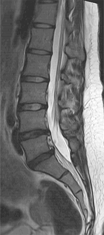
Fig. 1 Sagittal T2-weighted MR image (repetition time [TR] = 2750 ms, echo time [TE] = 104 ms) of the lumbar spine showing a mild degenerative bulging disc at L4-5 and L5-S1 levels.

Fig. 2 (a) Coronal T1-weighted MR image (TR = 691 ms, TE = 14 ms) revealing hypertrophy (asterisk) of the left piriformis muscle with an accessory belly (arrow) appearing at the inferior, proximal-third portion of the main piriformis muscle which coursed downward and laterally. (b) Axial T1-weighted MR image (TR = 709 ms, TE = 19 ms) showing the accessory belly (arrow) of the left piriformis muscle attaching to the greater trochanter, inferior to the insertion of the main piriformis muscle. (c) Sagittal T2-weighted MR image (TR = 3992 ms, TE = 100 ms) showing the accessory belly (arrowhead) at the inferior aspect of the left piriformis muscle along with thickening of the infrapiriformis portion of the left sciatic nerve (arrow). (d) Axial fat-suppressed T2-weighted MR image (TR = 4385 ms, TE = 70 ms) showing increased size and increased signal intensity of the left sciatic nerve (arrow). (e) Oblique coronal three-dimensional maximum intensity projection image obtained from fat-suppressed T2-weighted fast field echo (TR = 8 ms, TE = 4 ms, flap angle = 30°) clearly showing an abnormally thick left sciatic nerve (arrows) inferior to the hypertrophic left piriformis muscle (asterisk). (f) Coronal diffusion-weighted image with b value of 800 s/mm2 clearly demonstrating an abnormally thick left sciatic nerve (arrows) with diffusion restriction. (g, h) Axial T1-weighted (TR = 709 ms, TE = 19 ms) and axial fat-suppressed T2-weighted MR images (TR = 4385 ms, TE = 70 ms) showing no abnormal signal intensity suggesting denervation of left biceps femoris, semitendinosus, semimembranosus, and adductor magnus that are innervated by the left sciatic nerve.

Fig. 3 Drawings illustrating the six types of the Beaton and Anson's classification and our case regarding anatomical variation between the piriformis muscle and he sciatic nerve. Colored red and yellow indicate piriformis muscles and sciatic nerves (and its divisions), respectively. (a) Type A, undivided sciatic nerve exits the pelvis below the piriformis muscle. (b) Type B, sciatic nerve divides in the pelvis, common peroneal nerve pierces the piriformis muscle, and tibial nerve courses deep to the piriformis muscle. (c) Type C, sciatic nerve divides in the pelvis, common peroneal nerve courses over the piriformis muscle, and tibial nerve courses deep to the piriformis muscle. (d) Type D, undivided sciatic nerve exits the pelvis and pierces the piriformis muscle. (e) Type E, sciatic nerve divides in the pelvis, common peroneal nerve courses over the piriformis muscle, and tibial nerve pierces the piriformis muscle. (f) Type F, undivided sciatic nerve exits the pelvis and courses over the piriformis muscle. (g) Our case, accessory belly appears at the inferior aspect and proximal third portion of the main piriformis muscle, coursing laterally and inserting into the greater trochanter separately. Undivided sciatic nerve exits the pelvis below the main piriformis muscle and over the accessory belly.
Reference
-
1. Lee EY, Margherita AJ, Gierada DS, Narra VR. MRI of piriformis syndrome. AJR Am J Roentgenol. 2004; 183:63–64.
Article2. Petchprapa CN, Rosenberg ZS, Sconfienza LM, Cavalcanti CF, Vieira RL, Zember JS. MR imaging of entrapment neuropathies of the lower extremity. Part 1. The pelvis and hip. Radiographics. 2010; 30:983–100.3. Beaton LE, Anson BJ. The relation of the sciatic nerve and its subdivisions to the piriformis muscle. Anat Rec. 1937; 70:1–5.4. Sen A, Rajesh S. Accessory piriformis muscle: an easily identifiable cause of piriformis syndrome on magnetic resonance imaging. Neurol India. 2011; 59:769–771.
Article5. Yadav Y, Mehta V, Roy S, Suri R, Rath G. Superior gluteal nerve entrapment between two bellies of piriformis muscle. IJAV. 2010; 3:203–204.6. Polesello GC, Queiroz MC, Linhares JPT, Amaral DT, Ono NK. Anatomical variation of piriformis muscle as a cause of deep gluteal pain: diagnosis using MR neurography and treatment. Rev Bras Ortop. 2013; 48:114–117.
Article7. Ravindranath Y, Manjunath KY, Ravindranath R. Accessory origin of the piriformis muscle. Singapore Med J. 2008; 49:e217–e218.8. Natsis K, Totlis T, Konstantinidis GA, Paraskevas G, Piagkou M, Koebke J. Anatomical variations between the sciatic nerve and the piriformis muscle: a contribution to surgical anatomy in piriformis syndrome. Surg Radiol Anat. 2014; 36:273–280.
Article9. Yeoman W. The relation of arthritis of the sacro-iliac joint to sciatica, with an analysis of 100 cases. Lancet. 1928; 2:1119–1122.
Article
- Full Text Links
-
- Actions
-
Cited
- CITED
-
- Close
- Share
-
- Similar articles
-
- Post-radiation Piriformis Syndrome in a Cervical Cancer Patient: A Case Report
- Leg Weakness Caused by Bilateral Piriformis Syndrome: A Case Report
- Piriformis Syndrome (Sciatic Nerve Entrapment) Associated With Type C Sciatic Nerve Variation: A Report of Two Cases and Literature Review
- Gait Improvement after Botulinum Toxin Injection in a Patient with Piriformis Muscle Syndrome
- Looking beyond Piriformis Syndrome: Is It Really the Piriformis?
