J Dent Rehabil Appl Sci.
2019 Jun;35(2):90-97. 10.14368/jdras.2019.35.2.90.
Relation of head posture and occlusal contact area using photo occlusion analysis
- Affiliations
-
- 1Department of Prosthodontics and Research Institute of Oral Science, College of Dentistry, Gangneung-Wonju National University, Gangneung, Republic of Korea. doctorcj@gwnu.ac.kr
- KMID: 2451756
- DOI: http://doi.org/10.14368/jdras.2019.35.2.90
Abstract
- PURPOSE
Previous studies related with occlusal contact area were limited that interocclusal thickness level or the method of measurement has not been accurate in measuring. The purpose of this study was to investigate the relation between head posture and occlusal contact area using photo occlusion analysis.
MATERIALS AND METHODS
54 subjects with complete dentition (44 men, 10 women / 23 to 33 years of age) were included. To identify the relationship between head posture and occlusal contact area, subjects took interocclusal record in maximal intercuspal position with three different positions(supine position (0°) / inclined position (45°) / upright position (90°)) on the dental unit chair. Occlusal contact area was analyzed using photo occlusion analysis. Statistical analyses were performed with SPSS ver.25.0 at 95% confidence interval.
RESULTS
Head posture has no significant effect on the changes of occlusal contact area (P > 0.05).
CONCLUSION
When interocclusal relation is stable, head posture does not change a interocclusal record because head posture has no significant effect on occlusal contact area. Analysis of occlusal contact area using photo occlsion analysis device is useful due to its material property and simplicity.
Figure
-

Fig. 1 Head postures. (A) Supine position (0°, group S), (B) Inclined position (45°, group I), (C) Upright position (90°, group U).
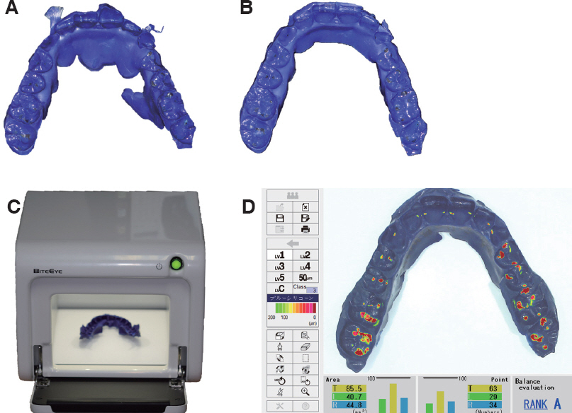
Fig. 2 Interocclusal record was modified to analyze. (A) Before modification, (B) After modification, (C) BiteEye® device, (D) Analysis of interocclusal record.
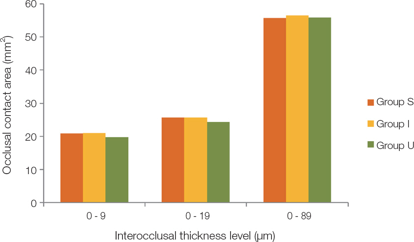
Fig. 3 Occlusal contact area according to head postures with each interocclusal thickness level.No significant differences were shown between group S, I and U.
Reference
-
References
1. Iwase Y, Saitoh I, Okamoto A, Nakakura-Ohshima K, Inada E, Yamada C, Takemoto Y, Yamasaki Y, Hayasaki H. Do occlusal contact areas of maximum closing position during gum chewing and intercuspal position coincide? Arch Oral Biol. 2011; 56:1616–23. DOI: 10.1016/j.archoralbio.2011.07.004. PMID: 21855855.2. English JD, Buschang PH, Throckmorton GS. Does malocclusion affect masticatory performance? Angle Orthod. 2002; 72:21–7. DOI: 10.1043/0003-3219(2002)072<0021:DMAMP>2.0.CO;2. PMID: 11843269.3. Wang YL, Cheng J, Chen YM, Yip KH, Smales RJ, Yin XM. Patterns and forces of occlusal contacts during lateral excursions recorded by the T-Scan II system in young Chinese adults with normal occlusions. J Oral Rehabil. 2011; 38:571–8. DOI: 10.1111/j.1365-2842.2010.02194.x. PMID: 21294761.4. Wilding RJ. The association between chewing efficiency and occlusal contact area in man. Arch Oral Biol. 1993; 38:589–96. DOI: 10.1016/0003-9969(93)90124-5. PMID: 8368956.5. Owens S, Buschang PH, Throckmorton GS, Palmer L, English J. Masticatory performance and areas of occlusal contact and near contact in subjects with normal occlusion and malocclusion. Am J Orthod Dentofacial Orthop. 2002; 121:602–9. DOI: 10.1067/mod.2002.122829. PMID: 12080313.6. Bae J, Son WS, Kim SS, Park SB, Kim YI. Comparison of masticatory efficiency according to Angle's classification of malocclusion. Korean J Orthod. 2017; 47:151–7. DOI: 10.4041/kjod.2017.47.3.151. PMID: 28523241. PMCID: PMC5432436.7. Lepley CR, Throckmorton GS, Ceen RF, Buschang PH. Relative contributions of occlusion, maximum bite force, and chewing cycle kinematics to masticatory performance. Am J Orthod Dentofacial Orthop. 2011; 139:606–13. PMID: 21536203. DOI: 10.1016/j.ajodo.2009.07.025.8. Bourdiol P, Mioche L. Correlations between functional and occlusal tooth-surface areas and food texture during natural chewing sequences in humans. Arch Oral Biol. 2000; 45:691–9. PMID: 10869481. DOI: 10.1016/S0003-9969(00)00027-3.9. Gupta S, Tarannum F, Gupta NK, Upadhyay M, Abdullah A. Effect of head posture on tooth contacts in dentate and complete denture wearers using computerized occlusal analysis system. J Indian Prosthodont Soc. 2017; 17:250–4. DOI: 10.4103/jips.jips_321_16. PMID: 28936038. PMCID: PMC5601498.10. Coelho MF, Cavalcanti Bd, Claro Neves AC, Joias RP, Rode S de M. Influence of dental chair backrest inclination on the registration of the mandibular position. J Prosthet Dent. 2015; 114:693–5. DOI: 10.1016/j.prosdent.2015.05.013. PMID: 26344187.11. Ohmure H, Miyawaki S, Nagata J, Ikeda K, Yamasaki K, Al-Kalaly A. Influence of forward head posture on condylar position. J Oral Rehabil. 2008; 35:795–800. PMID: 18808377. DOI: 10.1111/j.1365-2842.2007.01834.x.12. Ayub E, Glasheen-Way M, Kraus S. Head posture:a case study of the effects on the rest position of the mandible. J Orthop Sports Phys Ther. 1984; 5:179–83. DOI: 10.2519/jospt.1984.5.4.179. PMID: 18806411.13. Halperin GC, Halperin AR, Norling BK. Thickness, strength, and plastic deformation of occlusal registration strips. J Prosthet Dent. 1982; 48:575–8. DOI: 10.1016/0022-3913(82)90367-5. PMID: 6958865.14. Harper KA, Setchell DJ. The use of shimstock to assess occlusal contacts:a laboratory study. Int J Prosthodont. 2002; 15:347–52. PMID: 12170848.15. Millstein P, Maya A. An evaluation of occlusal contact marking indicators. A descriptive quantitative method. J Am Dent Assoc. 2001; 132:1280–6. DOI: 10.14219/jada.archive.2001.0373. PMID: 11665355.16. Ando K, Kurosawa M, Fuwa Y, Kondo T, Goto S. A study on measuring occlusal contact area using silicone impression materials:an application of this method to the bite force measurement system using the pressure-sensitive sheet. Dent Mater J. 2007; 26:898–905. DOI: 10.4012/dmj.26.898. PMID: 18203497.17. Millstein PL. An evaluation of occlusal indicator wax. J Prosthet Dent. 1985; 53:570–2. DOI: 10.1016/0022-3913(85)90651-1. PMID: 3858520.18. Agbaje JO, Casteele EV, Salem AS, Anumendem D, Shaheen E, Sun Y, Politis C. Assessment of occlusion with the T-Scan system in patients undergoing orthognathic surgery. Sci Rep. 2017; 7:5356. DOI: 10.1038/s41598-017-05788-x. PMID: 28706294. PMCID: PMC5509719.19. Park KW, Cho LR, Kim DG, Park CJ. Analysis of occlusal contacts using add-picture method. J Dent Rehabil Appl Sci. 2013; 29:45–58. DOI: 10.14368/jdras.2013.29.1.045.20. Kim CH, Kim DG, Ko KH, Huh YH, Cho LR, Park CJ. Considerations in the reliability of occlusal indicators and occlusal contact marks. J Dent Rehabil Appl Sci. 2018; 34:147–56. DOI: 10.14368/jdras.2018.34.3.147.21. Okada Y, Sato Y, Kitagawa N, Uchida K, Osawa T, Imamura Y, Terazawa M. Occlusal status of implant superstructures at mandibular first molar immediately after setting. Int J Implant Dent. 2015; 1:16. DOI: 10.1186/s40729-015-0016-0. PMID: 27747638. PMCID: PMC5005633.22. Nishimori H, Iida T, Kamiyama H, Komoda Y, Obara R, Uchida T, Kawara M, Komiyama O. Comparing the occlusal contact area of individual teeth during low-level clenching. J Oral Sci. 2017; 59:337–42. DOI: 10.2334/josnusd.16-0453. PMID: 28529279.23. Sato Y, Kitagawa N, Terazawa M, Osawa T, Imamura Y. Time-dependent changes in occlusal status in an implant-supported prosthesis at the mandibular first molar from prosthesis placement to 3 months after. Int J Prosthodont Restor Dent. 2018; 8:44–53. DOI: 10.5005/jp-journals-10019-1203.24. Imamura Y, Sato Y, Kitagawa N, Uchida K, Osawa T, Omori M, Okada Y. Influence of occlusal loading force on occlusal contacts in natural dentition. J Prosthodont Res. 2015; 59:113–20. DOI: 10.1016/j.jpor.2014.07.001. PMID: 25161117.25. Komiyama O, Obara R, Iida T, Asano T, Masuda M, Uchida T, De Laat A, Kawara M. Comparison of direct and indirect occlusal contact examinations with different clenching intensities. J Oral Rehabil. 2015; 42:185–91. DOI: 10.1111/joor.12242. PMID: 25270097.26. Obara R, Komiyama O, Iida T, De Laat A, Kawara M. Influence of the thickness of silicone registration material as a means for occlusal contact examination-an explorative study with different tooth clenching intensities. J Oral Rehabil. 2013; 40:834–43. DOI: 10.1111/joor.12088. PMID: 23889702.27. Terauchi R, Arai K, Tanaka M, Kawazoe T, Baba S. Effect of difference in occlusal contact area of mandibular free-end edentulous area implants on periodontal mechanosensitive threshold of adjacent premolars. Springerplus. 2015; 4:703. DOI: 10.1186/s40064-015-1497-2. PMID: 26609505. PMCID: PMC4648849.28. Uchida T, Komiyama O, Kubodera S, Okamoto Y, Iida T, Wakami M. The reliability of occlusal contact analysis using Blue Silicone®conformance inspection materials. J Jpn Soc TMJ. 2014; 26:93–9.29. Kubo H, Sato M, Kashiwagi K, Nakagawa S, Fujii T, Tanaka J, Tanaka M. Repeatability of the occlusal contact area measurements using a BiteEye®tooth contact analyzing device. J Osaka Odontol Soc. 2013; 76:83–8.
- Full Text Links
-
- Actions
-
Cited
- CITED
-
- Close
- Share
-
- Similar articles
-
- Effectiveness of clinical remounting improving balanced occlusion of complete dentures
- Changes of bite force and dynamic functional occlusion analysis after occlusal stabilization splint therapy in sleep bruxism patients: a pilot study
- Computerized analysis of occlusal contacts in bruxism patients treated with occlusal splint therapy
- Evaluation of occlusal strength using T-Scan Novus and Dental prescale II in dental prosthodontic treatments: A case report
- Comparisons of occlusal force according to occlusal relationship, skeletal pattern, age and gender in Koreans
