Comprehensive understandings in the shape of retromolar pad and its classification based on clinical application
- Affiliations
-
- 1Department of Prosthodontics and Research Institute of Oral Science, College of Dentistry, Gangneung-Wonju National University, Gangneung, Republic of Korea. lila@gwnu.ac.kr
- KMID: 2451753
- DOI: http://doi.org/10.14368/jdras.2019.35.2.64
Abstract
- Retromolar pad is one of anatomical landmarks to use clinically for determining of specific occlusal plane level in edentulous patients and is inherently a soft tissue mass; located at the distal end of mandible and should be covered with denture. The shapes of retromolar pad show variability because it might be inevitable to be resultant forms from healing and remodeling process after the extraction of last molars. Considerations would be focused on those resultant shapes and classification with anatomical relationships and clinical significances.
Keyword
Figure
-

Fig. 1 Bony structures of retromolar region. Retromolar triangle is located behind a third molar and retromolar fossa, an ovoid depression, is deeper, more laterally placed and larger than the retromolar triangle. Unlikely the retromolar triangle, it can be palpated. Retromolar fossa is bounded laterally by the external oblique ridge and its continuation in the anterior border of the ascending ramus; medially it is bounded by the temporal crest and at a lower level by the buccal border of the small retromolar triangle.
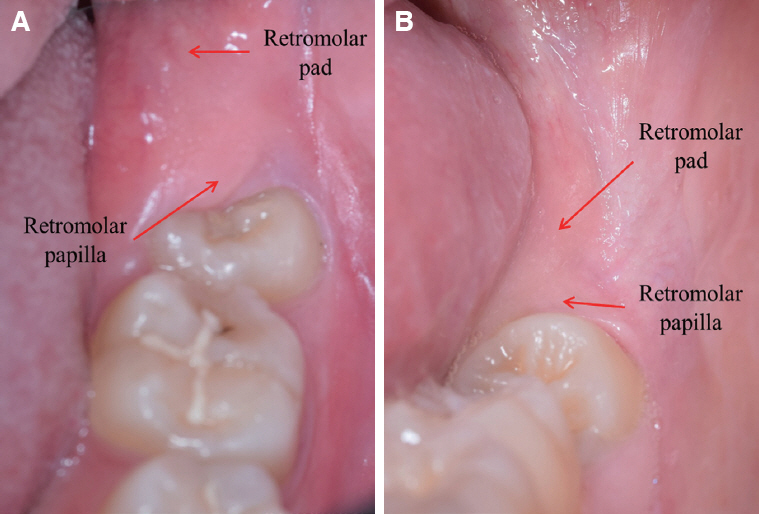
Fig. 2 Relationship between retromolar papilla and retromolar pad when a third molar is present. Directly overlying the base or mesial side of the bony retromolar triangle is the retromolar papilla, a small rolled or crescent-shaped elevation of the mucosa. Retromolar pad is posteriorly continuous with the pterygomandibular raphe. (A) The distal half of a third molar is partially covered with retromolar papilla. (B) Retromolar papilla is clearly located surrounding the distal surface of a third molar.
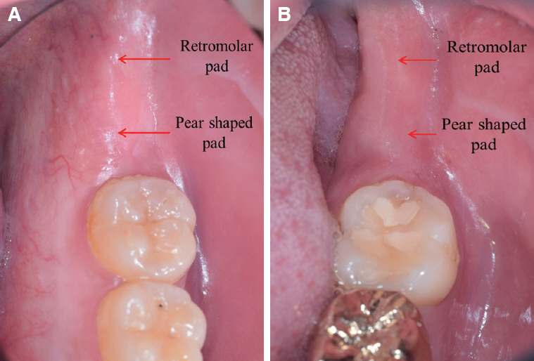
Fig. 3 Relationship between pear shaped pad and retromolar pad when a third molar is not present. After losing last molars, retromolar pad is anteriorly blended with a new elevation, so called, pear shaped pad. Pear shaped pad consists of fibrous tissue of the same kind as that forming the gingival mucosa, it is directly in line with the crest of the edentulous alveolar ridge, and it forms the distal termination of this residual structure. (A) An existed third molar was extracted and consecutively, soft tissue healing and bone remodeling process created the fusion with previously surrounding structures. (B) A third molar is congenitally missing. Relatively, smooth and long soft tissue continuity is shown behind a second molar.
Reference
-
References
1. Zarb G, Hobkirk JA, Eckert SE, Jacob RF. Prosthodontic treatment for edentulous patients. 13th ed. St. Louis: Elsevier Mosby;2013. p. 167–8.2. Craddock FW. Retromolar region of the mandible. J Am Dent Assoc. 1953; 47:453–5. DOI: 10.14219/jada.archive.1953.0195. PMID: 13108486.3. McCrorie JW, Hall DC. A histologic investigation of the retromolar and pear-shaped pad. Dent Pract Dent Rec. 1965; 15:237–9.4. Gruber H, Solar P, Ulm C. Maxillomandibular anatomy and patterns of resorption following atrophy. Watzek G, editor. Endosseous Implants. Scientific and Clinical Aspects;Chicago;Quintessence: 1993. p. 29–62.5. Wright CR. Evaluation of the factors necessary to develop stability in mandibular dentures. J Prosthet Dent. 1966; 16:414–30. DOI: 10.1016/0022-3913(66)90045-X.6. Pietrokovski J, Kaffe I, Arensburg B. Retromolar ridge in edentulous patients:clinical considerations. J Prosthodont. 2007; 16:502–6. DOI: 10.1111/j.1532-849X.2007.00225.x. PMID: 17672832.7. Ismail YH, Bowman JF. Position of the occlusal plane in natural and artificial teeth. J Prosthet Dent. 1968; 20:407–11. DOI: 10.1016/S0022-3913(68)80017-4. PMID: 4878987.8. Wright WH. Selection and arrangement of artificial teeth for complete prosthetic dentures. J Am Dent Assoc. 1936; 23:2291–307. DOI: 10.14219/jada.archive.1936.0353.9. Martone AL, Edwards LF. Anatomy of mouth and its related structures. J Prosthet Dent. 1962; 12:817–34. DOI: 10.1016/0022-3913(62)90003-3.10. Pendleton EC. The minute anatomy of the denture bearing area. J Am Dent Assoc. 1934; 21:488–504. DOI: 10.14219/jada.archive.1934.0072.11. Worthington P, Rubenstein JE. Problems associated with the atrophic mandible. Dent Clin North Am. 1998; 42:129–60. PMID: 9421674.12. Sinobad D. The position of occlusal plane in dentulous subjects with various skeletal jaw relations. J Oral Rehabil. 1988; 15:489–98. DOI: 10.1111/j.1365-2842.1988.tb00184.x. PMID: 3244056.13. Boucher CO. Swenson's complete dentures. 5th ed. St. Louis: CV Mosby;1964. p. 246–51.14. Hall WA. Important factors in adequate denture occlusion. J Prosthet Dent. 1958; 8:764–75. DOI: 10.1016/0022-3913(58)90096-9.15. Rahn AO, Heartwell CM. Textbook of complete dentures. 5th ed. Wolters Kluwer Co;1986. p. 352.16. Engelmeier RL. Complete denture esthetics. Dent Clin North Am. 1996; 40:71–84. PMID: 8635627.17. Gupta R, Aeran H, Singh SP. Relationship of anatomical landmarks with occlusal plane. J Indian Prosthodont Soc. 2009; 9:142–7. DOI: 10.4103/0972-4052.57083.18. Lunquist DO, Luther WW. Occlusal plane determination. J Prosthet Dent. 1970; 23:489–98. DOI: 10.1016/0022-3913(70)90198-8. PMID: 4908801.19. Shigli K, Chetal B, Jabade J. Validity of soft tissue landmarks in determining the occlusal plane. J Indian Prosthodont Soc. 2005; 5:139–45. DOI: 10.4103/0972-4052.17107.20. Yasaki M. Height of the occlusion rim and the interocclusal distance. J Prosthet Dent. 1961; 11:26–31. DOI: 10.1016/0022-3913(61)90106-8.21. Nagle RJ, Sears VH. Denture prosthetics. 2nd ed. St. Louis: CV Mosby;1962; 134.22. Ghosn CA, Zogheib C, Makzoume JE. Relationship between the occlusal plane corresponding to the lateral borders of the tongue and the ala-tragus line in edentulous patients. J Contemp Dent Pract. 2012; 13:590–4. DOI: 10.5005/jp-journals-10024-1192. PMID: 23250158.23. Nissan J, Barnea E, Zaltzer C, Cardash HS. Relationship between the occlusal plane determinants and craniofacial structures. J Oral Rehabil. 2003; 30:587–91. DOI: 10.1046/j.1365-2842.2003.01044.x. PMID: 12787455.24. Lopez FB, Suazo GI, Cantin LM, Sandoval MC. Biometrics study of the retromolar pad. Int J Odontostomat. 2008; 2:39–42.25. Sharma A, Deep A, Siwach A, Singh M, Bhargava A, Siwach R. Assessment and evaluation of anatomic variations of retromolar pad:a cross sectional study. J Clin Diag Res. 2016; 10:ZC143–5. DOI: 10.7860/JCDR/2016/19551.7880. PMID: 27437350. PMCID: PMCPMC4948526.
- Full Text Links
-
- Actions
-
Cited
- CITED
-
- Close
- Share
-
- Similar articles
-
- Three-dimensional morphometric study on the retromolar pad
- Morphometric Study of the Retromolar Triangle and Foramen in Korean Mandibles
- Pleomorphic Adenoma Which Occurred On The Left Retromolar Pad Area: A Case Report
- Clinical comparative Study Between Uncemented Anatomic Stem with and Without Lateral Porous Pad
- A study on the occlusal plane and the vertical dimension in Korean adults with natural dentition
