A review on the accuracy assessment methods of 3-dimensional digital dental models
- Affiliations
-
- 1Department of Prosthodontics and Dental Research Institute, School of Dentistry, Seoul National University, Seoul, Republic of Korea. limdds@snu.ac.kr
- 2Department of Periodontics, One-Stop Specialty Center, Seoul National University, Dental Hospital, Seoul, Republic of Korea.
- 3Clinical Translational Research Center for Dental Science, Seoul National University Dental Hospital, Seoul, Republic of Korea.
- KMID: 2451752
- DOI: http://doi.org/10.14368/jdras.2019.35.2.55
Abstract
- The aim of this article was to review various methods used to evaluate the accuracy of digital dental models. When evaluating the accuracy of digital models, the errors can be reduced by educating examiners and using artificial landmarks. The accuracy evaluation methods of digital dental models are divided into linear measurement, 2-dimensional cross-sectional analysis, and 3-dimensional best fit measurement. As the technology of scanners develops, many studies have been conducted to compare the accuracy of digital impression and conventional impression. According to improvement of scan technologies and development of 3-dimensional model analysis software, the ability to evaluate the accuracy of digital models is becoming more efficient. In this article, we describe the methods for evaluating the accuracy of a digital model and investigate effective accuracy analysis methods for each situation.
Keyword
Figure
-

Fig. 1 Accuracy, which includes Trueness and Precision, is represented by the bias and standard deviation of the Total error.

Fig. 2 Linear measurements made on each digital model. (A) Incisor to molar, (B) Intermolar width, (C) Incisor height.
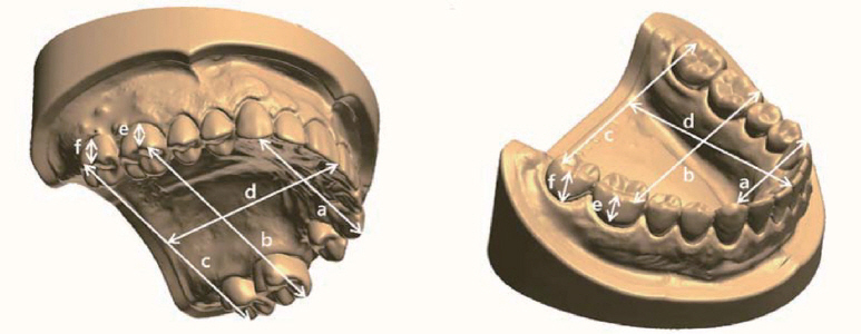
Fig. 3 Linear measurements at reference point of 3D model. a: inter-canine distance; b: inter-first molar distance; c:inter-second molar distance; d: arch length; e: first molar height; f: second molar height.
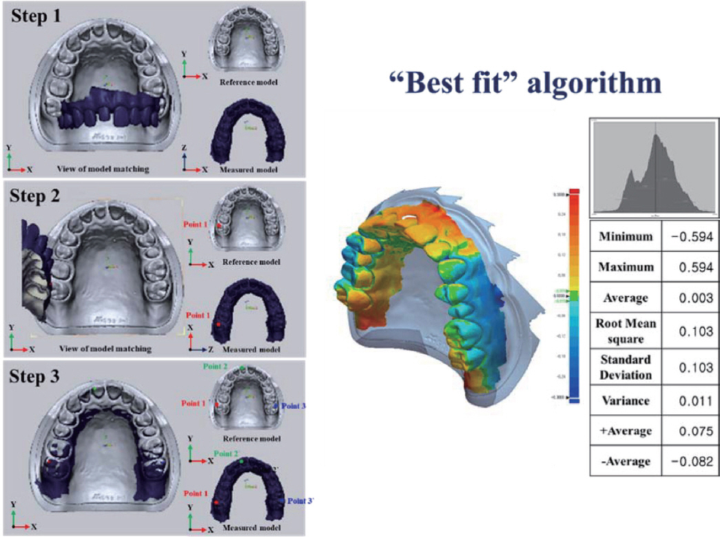
Fig. 4 Deviations after superimposition of scan datasets displayed in a color-coded using Best-Fit Alignment function in Geomagic Control XTM (3D systems, RockHill, USA).
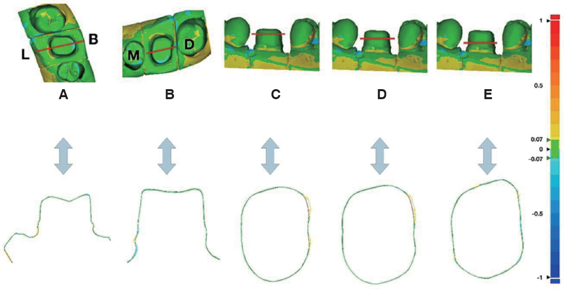
Fig. 5 2-dimensional cross-sectional analysis of abutment tooth. (A) Buccal-Lingual section, (B) Mesial-Distal section, (C) Transverse High section, (D) Transverse Middle section, (E) Transverse Low section. B: buccal; L: lingual; M: mesial; D: distal.17
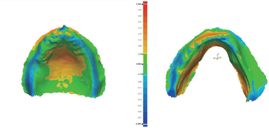
Fig. 6 Deviations after superimposition of edentulous scan datasets displayed in a color-coded difference image.
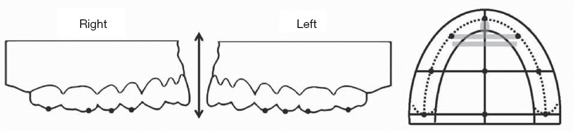
Fig. 7 Anatomical landmarks on a polished surface and a denture base surface.
Reference
-
References
1. Seelbach P, Brueckel C, Wostmann B. Accuracy of digital and conventional impression techniques and workflow. Clin Oral Investig. 2013; 17:1759–64. DOI: 10.1007/s00784-012-0864-4. PMID: 23086333.2. Ender A, Attin T, Mehl A. In vivo precision of conventional and digital methods of obtaining complete-arch dental impressions. J Prosthet Dent. 2016; 115:313–20. DOI: 10.1016/j.prosdent.2015.09.011. PMID: 26548890.3. Kim JH, Kim KB, Kim WC, Kim JH, Kim HY. Accuracy and precision of polyurethane dental arch models fabricated using a three-dimensional subtractive rapid prototyping method with an intraoral scanning technique. Korean J Orthod. 2014; 44:69–76. DOI: 10.4041/kjod.2014.44.2.69. PMID: 24696823. PMCID: PMCPMC3971128.4. Jemt T, Hjalmarsson L. In vitro measurements of precision of fit of implant-supported frameworks. A comparison between “virtual” and “physical” assessments of fit using two different techniques of measurements. Clin Implant Dent Relat Res. 2012; 14(Suppl 1):e175–82. DOI: 10.1111/j.1708-8208.2011.00416.x. PMID: 22171700.5. Gonzalez de Villaumbrosia P, Martinez-Rus F, Garcia-Orejas A, Salido MP, Pradies G. In vitro comparison of the accuracy (trueness and precision) of six extraoral dental scanners with different scanning technologies. J Prosthet Dent. 2016; 116:543–50. DOI: 10.1016/j.prosdent.2016.01.025. PMID: 27112413.6. Quimby ML, Vig KW, Rashid RG, Firestone AR. The accuracy and reliability of measurements made on computer-based digital models. Angle Orthod. 2004; 74:298–303. DOI: 10.1043/0003-3219(2004)074<0298:TAAROM>2.0.CO;2. PMID: 15264638.7. Menditto A, Patriarca M, Magnusson B. Understanding the meaning of accuracy, trueness and precision. Accredit Qual Assur. 2006; 12:45–7. DOI: 10.1007/s00769-006-0191-z.8. Mai HN, Lee KE, Ha JH, Lee DH. Effects of image and education on the precision of the measurement method for evaluating prosthesis misfit. J Prosthet Dent. 2018; 119:600–5. DOI: 10.1016/j.prosdent.2017.05.022. PMID: 28781069.9. Kim JE, Amelya A, Shin Y, Shim JS. Accuracy of intraoral digital impressions using an artificial landmark. J Prosthet Dent. 2017; 117:755–61. DOI: 10.1016/j.prosdent.2016.09.016. PMID: 27863856.10. Jiang T, Lee SM, Hou Y, Chang X, Hwang HS. Evaluation of digital dental models obtained from dental cone-beam computed tomography scan of alginate impressions. Korean J Orthod. 2016; 46:129–36. DOI: 10.4041/kjod.2016.46.3.129. PMID: 27226958. PMCID: PMCPMC4879315.11. Joo YH, Lee JH. Three dimensional accuracy analysis of dental stone casts fabricated using irreversible hydrocolloid impressions. J Dent Rehabil Appl Sci. 2015; 31:316–28. DOI: 10.14368/jdras.2015.31.4.316.12. Lee JH, Paik KS, Chang MS, Lee SP. An evaluation of validity of measurements using digital caliper and three-dimensional virtual dental models. Korean J Anat. 2004; 37:209–18.13. Guth JF, Keul C, Stimmelmayr M, Beuer F, Edelhoff D. Accuracy of digital models obtained by direct and indirect data capturing. Clin Oral Investig. 2013; 17:1201–8. DOI: 10.1007/s00784-012-0795-0. PMID: 22847854.14. Ender A, Mehl A. Accuracy of complete-arch dental impressions:a new method of measuring trueness and precision. J Prosthet Dent. 2013; 109:121–8. DOI: 10.1016/S0022-3913(13)60028-1. PMID: 23395338.15. Renne W, Ludlow M, Fryml J, Schurch Z, Mennito A, Kessler R, Lauer A. Evaluation of the accuracy of 7 digital scanners:an in vitro analysis based on 3-dimensional comparisons. J Prosthet Dent. 2017; 118:36–42. DOI: 10.1016/j.prosdent.2016.09.024. PMID: 28024822.16. Ender A, Zimmermann M, Attin T, Mehl A. In vivo precision of conventional and digital methods for obtaining quadrant dental impressions. Clin Oral Investig. 2016; 20:1495–504. DOI: 10.1007/s00784-015-1641-y. PMID: 26547869.17. Maeng J, Lim YJ, Kim B, Kim MJ, Kwon HB. A new approach to accuracy evaluation of single-tooth abutment using two-dimensional analysis in two intraoral scanners. Int J Environ Res Public Health. 2019; 16:E1021. DOI: 10.3390/ijerph16061021. PMID: 30897832. PMCID: PMCPMC6466129.18. Patzelt SB, Vonau S, Stampf S, Att W. Assessing the feasibility and accuracy of digitizing edentulous jaws. J Am Dent Assoc. 2013; 144:914–20. DOI: 10.14219/jada.archive.2013.0209. PMID: 23904578.19. Goodacre BJ, Goodacre CJ, Baba NZ, Kattadiyil MT. Comparison of denture base adaptation between CAD-CAM and conventional fabrication techniques. J Prosthet Dent. 2016; 116:249–56. DOI: 10.1016/j.prosdent.2016.02.017. PMID: 27112416.20. Norvell NG, Korioth TV, Cagna DR, Versluis A. Comparison of digital surface displacements of maxillary dentures based on noninvasive anatomic landmarks. J Prosthet Dent. 2018; 120:123–31. DOI: 10.1016/j.prosdent.2017.11.006. PMID: 29429839.
- Full Text Links
-
- Actions
-
Cited
- CITED
-
- Close
- Share
-
- Similar articles
-
- Clinical Validity of Tooth Size Measurements Obtained via Digital Methods with Intraoral Scanning
- Accuracy of conventional and digital mounting of dental models: A literature review
- Three dimensional accuracy analysis of dental stone casts fabricated using irreversible hydrocolloid impressions
- Comparison of digital models generated from three-dimensional optical scanner and cone beam computed tomography
- An Evaluation of Validity of Measurements using Digital Caliper and Three-dimensional Virtual Dental Models
