Obstet Gynecol Sci.
2019 Jul;62(4):273-279. 10.5468/ogs.2019.62.4.273.
The significance of sonographically thickened endometrium in asymptomatic postmenopausal women
- Affiliations
-
- 1Department of Reproductive Endocrinology and Infertility, University of Health Sciences, Ankara Etlik Zubeyde Hanım Women's Health Application and Research Center, Ankara, Turkey. runakara@gmail.com
- KMID: 2451657
- DOI: http://doi.org/10.5468/ogs.2019.62.4.273
Abstract
OBJECTIVE
To investigate the correlation between sonographic, hysteroscopic, and pathological findings in postmenopausal asymptomatic patients with sonographically thickened endometrium.
METHODS
The records of postmenopausal patients who attended the Menopause Outpatient Clinic of a tertiary women's hospital in Ankara, Turkey between January 1, 2012 and December 15, 2013 were retrieved. A total of 266 postmenopausal women without vaginal bleeding underwent hysteroscopic evaluation and endometrial sampling. Patients whose pathological records indicated an endometrial thickness equal to or greater than 6 mm (double layer) on transvaginal ultrasonography without any symptoms were included in the study.
RESULTS
The most frequently detected focal intrauterine lesions in asymptomatic women were endometrial polyps, which were diagnosed in 168 (63.1%) cases. Twenty-four (9%) patients were diagnosed as having simple hyperplasia, 4 (1%) atypical hyperplasia, and 8 (3%) endometrial adenocarcinoma. Two of the patients with adenocarcinoma were diagnosed based on endometrial polyps, and 6 cases showed endometrial hyperplasia on hysteroscopy, while histological examination showed endometrial carcinoma.
CONCLUSION
We suggest 10.5 mm as the cutoff value for endometrial thickness and recommend hysteroscopy following dilatation and curettage to increase diagnostic efficacy and provide definitive treatment in asymptomatic postmenopausal women with thickened endometrium.
Keyword
MeSH Terms
Figure
-
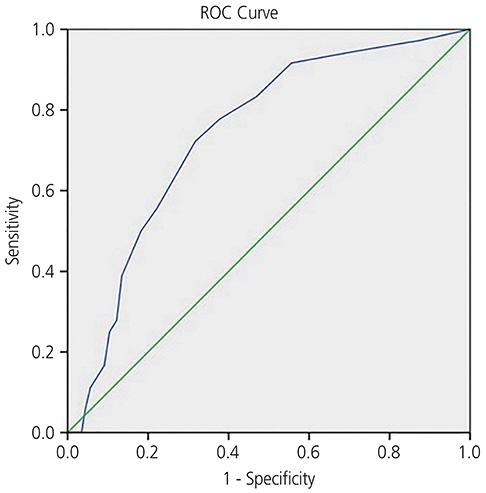
Fig. 1 Receiver operating characteristic (ROC) curves analysing the optimal cut-off value for premalignant and malignant lesions. Endometrial thickness in premalignant and malignant lesion prediction (area under the receiver operating characteristic curve=0.740; P=0.01; 95% confidence interval, 0.66–0.81).
Reference
-
1. Jemal A, Siegel R, Ward E, Murray T, Xu J, Thun MJ. Cancer statistics, 2007. CA Cancer J Clin. 2007; 57:43–66.
Article2. Kanat-Pektas M, Gungor T, Mollamahmutoglu L. The evaluation of endometrial tumors by transvaginal and Doppler ultrasonography. Arch Gynecol Obstet. 2008; 277:495–499.
Article3. van Hanegem N, Breijer MC, Khan KS, Clark TJ, Burger MP, Mol BW, et al. Diagnostic evaluation of the endometrium in postmenopausal bleeding: an evidence-based approach. Maturitas. 2011; 68:155–164.
Article4. Wong AS, Lao TT, Cheung CW, Yeung SW, Fan HL, Ng PS, et al. Reappraisal of endometrial thickness for the detection of endometrial cancer in postmenopausal bleeding: a retrospective cohort study. BJOG. 2016; 123:439–446.
Article5. Kasraeian M, Asadi N, Ghaffarpasand F, Karimi AA. Value of transvaginal ultrasonography in endometrial evaluation of non-bleeding postmenopausal women. Climacteric. 2011; 14:126–131.
Article6. Emanuel MH, Verdel MJ, Wamsteker K, Lammes FB. A prospective comparison of transvaginal ultrasonography and diagnostic hysteroscopy in the evaluation of patients with abnormal uterine bleeding: clinical implications. Am J Obstet Gynecol. 1995; 172:547–552.
Article7. Smith RA, Cokkinides V, Eyre HJ. American Cancer Society guidelines for the early detection of cancer, 2006. CA Cancer J Clin. 2006; 56:11–25.
Article8. Brandner P, Gnirs J, Neis KJ, Hettenbach A, Schmidt W. Value of vaginal ultrasonography in noninvasive assessment of the endometrium of the postmenopausal uterus. Geburtshilfe Frauenheilkd. 1991; 51:734–740.9. Osmers R, Völksen M, Schauer A. Vaginosonography for early detection of endometrial carcinoma? Lancet. 1990; 335:1569–1571.
Article10. Kim MJ, Kim JJ, Kim SM. Endometrial evaluation with transvaginal ultrasonography for the screening of endometrial hyperplasia or cancer in premenopausal and perimenopausal women. Obstet Gynecol Sci. 2016; 59:192–200.
Article11. Schmidt T, Breidenbach M, Nawroth F, Mallmann P, Beyer IM, Fleisch MC, et al. Hysteroscopy for asymptomatic postmenopausal women with sonographically thickened endometrium. Maturitas. 2009; 62:176–178.
Article12. Lev-Sagie A, Hamani Y, Imbar T, Hurwitz A, Lavy Y. The significance of intrauterine lesions detected by ultrasound in asymptomatic postmenopausal patients. BJOG. 2005; 112:379–381.
Article13. Weigel M, Friese K, Strittmatter HJ, Melchert F. Ultrasound assessment of the postmenopausal endometrium. Is measuring thickness adequate? Ultraschall Med. 1994; 15:117–121.14. Wethington SL, Herzog TJ, Burke WM, Sun X, Lerner JP, Lewin SN, et al. Risk and predictors of malignancy in women with endometrial polyps. Ann Surg Oncol. 2011; 18:3819–3823.
Article15. Antunes A Jr, Costa-Paiva L, Arthuso M, Costa JV, Pinto-Neto AM. Endometrial polyps in pre- and postmenopausal women: factors associated with malignancy. Maturitas. 2007; 57:415–421.
Article16. Menon U, Gentry-Maharaj A, Ryan A, Sharma A, Burnell M, Hallett R, et al. Recruitment to multicentre trials--lessons from UKCTOCS: descriptive study. BMJ. 2008; 337:a2079.
Article17. Worley MJ Jr, Dean KL, Lin SN, Caputo TA, Post RC. The significance of a thickened endometrial echo in asymptomatic postmenopausal patients. Maturitas. 2011; 68:179–181.
Article18. Saatli B, Yildirim N, Olgan S, Koyuncuoglu M, Emekci O, Saygılı U. The role of endometrial thickness for detecting endometrial pathologies in asymptomatic postmenopausal women. Aust N Z J Obstet Gynaecol. 2014; 54:36–40.
Article19. Breijer MC, Peeters JA, Opmeer BC, Clark TJ, Verheijen RH, Mol BW, et al. Capacity of endometrial thickness measurement to diagnose endometrial carcinoma in asymptomatic postmenopausal women: a systematic review and meta-analysis. Ultrasound Obstet Gynecol. 2012; 40:621–629.
Article20. Gambacciani M, Monteleone P, Ciaponi M, Sacco A, Genazzani AR. Clinical usefulness of endometrial screening by ultrasound in asymptomatic postmenopausal women. Maturitas. 2004; 48:421–424.
Article21. Yasa C, Dural O, Bastu E, Ugurlucan FG, Nehir A, İyibozkurt AC. Evaluation of the diagnostic role of transvaginal ultrasound measurements of endometrial thickness to detect endometrial malignancy in asymptomatic postmenopausal women. Arch Gynecol Obstet. 2016; 294:311–316.
Article22. Smith-Bindman R, Weiss E, Feldstein V. How thick is too thick? When endometrial thickness should prompt biopsy in postmenopausal women without vaginal bleeding. Ultrasound Obstet Gynecol. 2004; 24:558–565.
Article23. Widrich T, Bradley LD, Mitchinson AR, Collins RL. Comparison of saline infusion sonography with office hysteroscopy for the evaluation of the endometrium. Am J Obstet Gynecol. 1996; 174:1327–1334.
Article24. Pettersson B, Adami HO, Lindgren A, Hesselius I. Endometrial polyps and hyperplasia as risk factors for endometrial carcinoma. A case-control study of curettage specimens. Acta Obstet Gynecol Scand. 1985; 64:653–659.
Article
- Full Text Links
-
- Actions
-
Cited
- CITED
-
- Close
- Share
-
- Similar articles
-
- The effects of Raloxifene therapy on endometrium in postmenopausal women
- Analysis of Abnormally Thickened Endometrial Patterns on Trans vaginal Sonography
- The effect of adjuvant hormonal therapy on the endometrium and ovary of breast cancer patients
- The Usefulness of Vaginal Sonography in Evaluation of Endometrium in Postmenopausal Women
- The Correlation between the Endometrial Cancer and Postmenopausal Uterine Bleeding
