Portal Hypertension of a Delayed Onset Following Liver Abscesses in a 12-Month-Old Infant: A Case Report and Review of the Literature
- Affiliations
-
- 1Department of Pediatrics, College of Medicine, Imam Abdulrahman Bin Faisal University, Dammam, Saudi Arabia. Faisal.alqurashi@yahoo.com
- 2Department of Radiology, College of Medicine, Imam Abdulrahman Bin Faisal University, Dammam, Saudi Arabia.
- KMID: 2451581
- DOI: http://doi.org/10.5223/pghn.2019.22.4.400
Abstract
- We report a 12-month-old female infant who had a history of neonatal sepsis with liver micro-abscesses that resolved with intravenous antibiotics during neonatal period. During her neonatal admission period, no umbilical vein catheter was inserted. Also, she did not undergo any abdominal surgeries or had a postnatal history of necrotizing enterocolitis. However, the child developed upper gastrointestinal bleeding in form of hematemesis and melena secondary to esophageal varices at the age of 12 months with an extra-hepatic portal vein obstruction with cavernous transformation and portal hypertension subsequently. The child underwent a successful endoscopic injection sclerotherapy. She is now 20-month-old and has portal hypertension but otherwise asymptomatic. We are proposing the possibility of a delayed-onset portal hypertension as a complication of liver abscess and neonatal sepsis.
Keyword
MeSH Terms
Figure
-
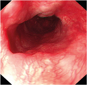
Fig. 1 Endoscopic view of grade II esophageal varices. An endoscopic view of the two esophageal varices, both of grade II, first one at 10 o'clock position, and second one was seen at 12 o'clock position.
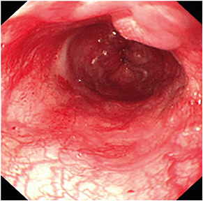
Fig. 2 Endoscopic view of grade II esophageal varices. An endoscopic view of the second esophageal varices at 12 o'clock position which extends from the level of 15 cm to the gastroesophageal junction at the level of 21 cm.
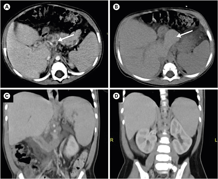
Fig. 3 CT angiogram. (A) Contrast enhanced axial CT scan of the abdomen showing dilated tortious vessels at the porta-hepatis and peripancreatic areas (arrow), in keeping with cavernous transformation of portal vein caused by portal hypertension. (B) Non-enhanced axial CT scan of the abdomen showing hypertrophy of the caudate lobe (arrow). (C) Contrast enhanced coronal CT scan of the abdomen showing splenomegaly and (D) minimal ascites at the sub-hepatic region. CT: computed tomography.
Reference
-
1. Garel D, Wood C, Pariente D, Dommergues JP. Portal system obstruction of delayed onset following neonatal Staphylococcus aureus infection. Arch Fr Pediatr. 1989; 46:41–43.2. Vade A, Sajous C, Anderson B, Challapalli M. Neonatal hepatic abscess. Comput Med Imaging Graph. 1998; 22:357–359.
Article3. Sethi SK, Dewan P, Faridi MM, Aggarwal A, Upreti L. Liver abscess, portal vein thrombosis and cavernoma formation following umbilical vein catherisation in two neonates. Trop Gastroenterol. 2007; 28:79–80.4. Shah I, Bhatnagar S. Liver abscess in a newborn leading to portal vein thrombosis. Indian J Pediatr. 2009; 76:1268–1269.
Article5. Sherwani P, Vire A, Anand R, Jajoo M. Umbilical venous catheterization gone wrong: hepatic complications. Indian J Radiol Imaging. 2016; 26:40–43.
Article6. Aggarwal S, Mathur NB, Garg A. Portal vein thrombosis complicating neonatal hepatic abscess. Indian Pediatr. 2003; 40:997–1001.7. Tan NW, Sriram B, Tan-Kendrick AP, Rajadurai VS. Neonatal hepatic abscess in preterm infants: a rare entity? Ann Acad Med Singapore. 2005; 34:558–564.8. Doerr CA, Demmler GJ, Garcia-Prats JA, Brandt ML. Solitary pyogenic liver abscess in neonates: report of three cases and review of the literature. Pediatr Infect Dis J. 1994; 13:64–69.9. Chawla YK, Bodh V. Portal vein thrombosis. J Clin Exp Hepatol. 2015; 5:22–40.
Article10. Poddar U, Thapa BR, Rao KL, Singh K. Etiological spectrum of esophageal varices due to portal hypertension in Indian children: is it different from the West? J Gastroenterol Hepatol. 2008; 23:1354–1357.
Article11. Valla DC, Condat B, Lebrec D. Spectrum of portal vein thrombosis in the West. J Gastroenterol Hepatol. 2002; 17:Suppl 3. S224–S227.
Article12. Rangari M, Gupta R, Jain M, Malhotra V, Sarin SK. Hepatic dysfunction in patients with extrahepatic portal venous obstruction. Liver Int. 2003; 23:434–439.
Article13. Arora NK, Lodha R, Gulati S, Gupta AK, Mathur P, Joshi MS, et al. Portal hypertension in north Indian children. Indian J Pediatr. 1998; 65:585–591.14. El-Karaksy HM, El-Koofy N, Mohsen N, Helmy H, Nabil N, El-Shabrawi M. Extrahepatic portal vein obstruction in Egyptian children. J Pediatr Gastroenterol Nutr. 2015; 60:105–109.
Article15. Blei AT, Groszmann RJ. Vasopressin and Vasoconstrictors. In : Shepherd AP, Granger DN, editors. The Physiology of the Intestinal Microcirculation. New York (NY): Raven Press;1984. p. 377.16. Bloom SR, Polak JM. Somatostatin. Br Med J (Clin Res Ed). 1987; 295:288–290.
Article17. Imperiale TF, Teran JC, McCullough AJ. A meta-analysis of somatostatin versus vasopressin in the management of acute esophageal variceal hemorrhage. Gastroenterology. 1995; 109:1289–1294.
Article18. Zargar SA, Javid G, Khan BA, Yattoo GN, Shah AH, Gulzar GM, et al. Endoscopic ligation compared with sclerotherapy for bleeding esophageal varices in children with extrahepatic portal venous obstruction. Hepatology. 2002; 36:666–672.
Article19. Kim SJ, Kim KM. Recent trends in the endoscopic management of variceal bleeding in children. Pediatr Gastroenterol Hepatol Nutr. 2013; 16:1–9.
Article20. Bari K, Garcia-Tsao G. Treatment of portal hypertension. World J Gastroenterol. 2012; 18:1166–1175.
Article
- Full Text Links
-
- Actions
-
Cited
- CITED
-
- Close
- Share
-
- Similar articles
-
- A Case of Pulmonary Hypertension Associated with Portal Hypertension
- Hepatic venous pressure gradient: clinical use in chronic liver disease
- Congenital Absence of the Portal Vein Presenting as Pulmonary Hypertension
- Preduodenal Portal Vein Associated with Duodenal Obstruction: A case report
- Portal vein fenestration: a case report of an unusual portal vein developmental anomaly
