Pediatr Gastroenterol Hepatol Nutr.
2019 Jul;22(4):350-357. 10.5223/pghn.2019.22.4.350.
Tissue Transglutaminase Antibody and Its Association with Duodenal Biopsy in Diagnosis of Pediatric Celiac Disease
- Affiliations
-
- 1Pediatric Intensive Care Unit, Great Ormond Street Hospital, London, United Kingdom. akunurishalini@gmail.com
- 2Department of Pediatrics, Lady Harding Medical College, New Delhi, India.
- 3Department of Pediatrics, Government BDM Hospital, Jaipur, India.
- 4Department of Pediatrics, SMS Medical College, Jaipur, India.
- KMID: 2451575
- DOI: http://doi.org/10.5223/pghn.2019.22.4.350
Abstract
- PURPOSE
This study aimed to evaluate a possible association between the anti-tissue transglutaminase antibody (anti-tTG) titer and stage of duodenal mucosal damage and assess a possible cut-off value of anti-tTG at which celiac disease (CD) may be diagnosed in children in conjunction with clinical judgment.
METHODS
This observational study was conducted at a gastroenterology clinic in a tertiary hospital from April 2012 to May 2013. Seventy children between 6-months and 18-years-old with suspected CD underwent celiac serology and duodenal biopsy. Statistical analyses were done using SPSS 16. Diagnostic test values were determined for comparing the anti-tTG titer with duodenal biopsy. An analysis of variance and Tukey-Kramer tests were performed for comparing the means between groups. A receiver operating characteristics curve was plotted to determine various cut-off values of anti-tTG.
RESULTS
The mean antibody titer increased with severity of Marsh staging (p<0.001). An immunoglobulin (Ig) A-tTG value at 115 AU/mL had 76% sensitivity and 100% specificity with a 100% positive predictive value (PPV) and 17% negative predictive value (NPV) for diagnosis of CD (p<0.001, 95% confidence interval [CI], 0.75-1).
CONCLUSION
There is an association between the anti-tTG titer and stage of duodenal mucosal injury in children with CD. An anti-tTG value of 115 AU/mL (6.4 times the upper normal limit) had 76% sensitivity, 100% specificity, with a 100% PPV, and 17% NPV for diagnosing CD (95% CI, 0.75-1). This cut-off may be used in combination with clinical judgment to diagnose CD.
Keyword
MeSH Terms
Figure
-

Fig. 1 Flow diagram. tTG: tissue transglutaminase enzyme.
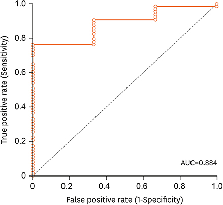
Fig. 2 ROC curve for anti-tTG titers and prediction of CD. ROC: receiver operated characteristics, tTG: tissue transglutaminase enzyme, AUC: area under the curve, CD: celiac disease.

Fig. 3 Correlation between age and anti-tTG titers. tTG: tissue transglutaminase enzyme.
Reference
-
1. Donaldson MR, Book LS, Leiferman KM, Zone JJ, Neuhausen SL. Strongly positive tissue transglutaminase antibodies are associated with Marsh 3 histopathology in adult and pediatric celiac disease. J Clin Gastroenterol. 2008; 42:256–260.
Article2. Kalhan S, Joseph P, Sharma S, Dubey S, Dudani S, Dixit M. Comparative study of histopathological Marsh grading with clinical and serological parameters in celiac iceberg of north India. Indian J Pathol Microbiol. 2011; 54:279–283.
Article3. Parizade M, Bujanover Y, Weiss B, Nachmias V, Shainberg B. Performance of serology assays for diagnosing celiac disease in a clinical setting. Clin Vaccine Immunol. 2009; 16:1576–1582.
Article4. Barker CC, Mitton C, Jevon G, Mock T. Can tissue transglutaminase antibody titers replace small-bowel biopsy to diagnose celiac disease in select pediatric populations? Pediatrics. 2005; 115:1341–1346.
Article5. Sugai E, Vázquez H, Nachman F, Moreno ML, Mazure R, Smecuol E, et al. Accuracy of testing for antibodies to synthetic gliadin-related peptides in celiac disease. Clin Gastroenterol Hepatol. 2006; 4:1112–1117.
Article6. Vivas S, Ruiz de Morales JG, Riestra S, Arias L, Fuentes D, Alvarez N, et al. Duodenal biopsy may be avoided when high transglutaminase antibody titers are present. World J Gastroenterol. 2009; 15:4775–4780.
Article7. Oberhuber G, Granditsch G, Vogelsang H. The histopathology of coeliac disease: time for a standardized report scheme for pathologists. Eur J Gastroenterol Hepatol. 1999; 11:1185–1194.8. Zanini B, Magni A, Caselani F, Lanzarotto F, Carabellese N, Villanacci V, et al. High tissue-transglutaminase antibody level predicts small intestinal villous atrophy in adult patients at high risk of celiac disease. Dig Liver Dis. 2012; 44:280–285.
Article9. Alessio MG, Tonutti E, Brusca I, Radice A, Licini L, Sonzogni A, et al. Correlation between IgA tissue transglutaminase antibody ratio and histological finding in celiac disease. J Pediatr Gastroenterol Nutr. 2012; 55:44–49.
Article10. Rahmati A, Shakeri R, Sohrabi M, Alipour A, Boghratian A, Setareh M, et al. Correlation of tissue transglutaminase antibody with duodenal histologic marsh grading. Middle East J Dig Dis. 2014; 6:131–136.11. Paul SP, Harries SL, Basude D. Barriers to implementing the revised ESPGHAN guidelines for coeliac disease in children: a cross-sectional survey of coeliac screen reporting in laboratories in England. Arch Dis Child. 2017; 102:942–946.
Article12. Husby S, Koletzko S, Korponay-Szabó IR, Mearin ML, Phillips A, Shamir R, et al. European Society for Pediatric Gastroenterology, Hepatology, and Nutrition guidelines for the diagnosis of coeliac disease. J Pediatr Gastroenterol Nutr. 2012; 54:136–160.
Article
- Full Text Links
-
- Actions
-
Cited
- CITED
-
- Close
- Share
-
- Similar articles
-
- Association between Celiac Disease and Intussusceptions in Children: Two Case Reports and Literature Review
- Diagnostic Value of Immunoglobulin G Anti-Deamidated Gliadin Peptide Antibody for Diagnosis of Pediatric Celiac Disease: A Study from Shiraz, Iran
- Clinical Significance of Anti-tissue Transglutaminase as a Screening Method for Celiac Disease in a Korean Population
- Can a Synbiotic Supplementation Contribute to Decreasing Anti-Tissue Transglutaminase Levels in Children with Potential Celiac Disease?
- Coeliac Disease-Associated Antibodies in Psoriasis
